
Virtual Living Cell
An animal cell and a plant cell that are alive. Tap any organelle to see what it is and what it does, then watch the cell work: respiration making ATP, photosynthesis, the insulin gene becoming a protein, and water moving in and out.
Experiment
HealthyTypical animal cell0:00
Your cell
ATP—
ATP made—
Aerobic—
Glucose used—
Oxygen—
Photosynthesis—
Water—
Health—
Organelle
Surroundings
Activity & poisons
Lab
View & time
What the cell is doing
Lab notebook
Every measurement you record, and every big event in your cell (switching to anaerobic respiration, bursting, plasmolysis, running out of energy), is written here with its conditions. Export a CSV, or turn the session into a PDF lab report.
| # | Cell | Conditions | Measurement | Result |
|---|
Concept library
Click "New problem" to begin.
Click "Start quiz" for 5 questions on organelles, animal and plant cells, respiration, photosynthesis, proteins and osmosis.
User Guide: Virtual Living Cell
1 Overview
Simulate shows a living cell. Choose Animal or Plant and a cell type in the top bar, tap any part of the cell to find out about it, and press a Show me button on the cell to watch respiration, photosynthesis, protein synthesis, osmosis, membrane transport or phagocytosis step by step. Change the cell's surroundings (oxygen, glucose, temperature, light, carbon dioxide, the outside solution) and poison it, and see what happens to its ATP. Experiment has seven labs that end in a PDF lab report, and Quiz has a practice quiz with worked solutions and a 5-question quiz.
2 The cells
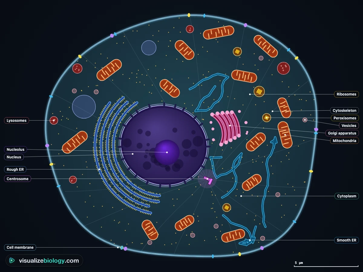
- Animal: a typical animal cell (as in a textbook diagram), a liver cell (many mitochondria, smooth ER, peroxisomes, glycogen and often two nuclei), a pancreatic β-cell (rough ER, Golgi apparatus and insulin granules) and a macrophage (lysosomes, for engulfing bacteria).
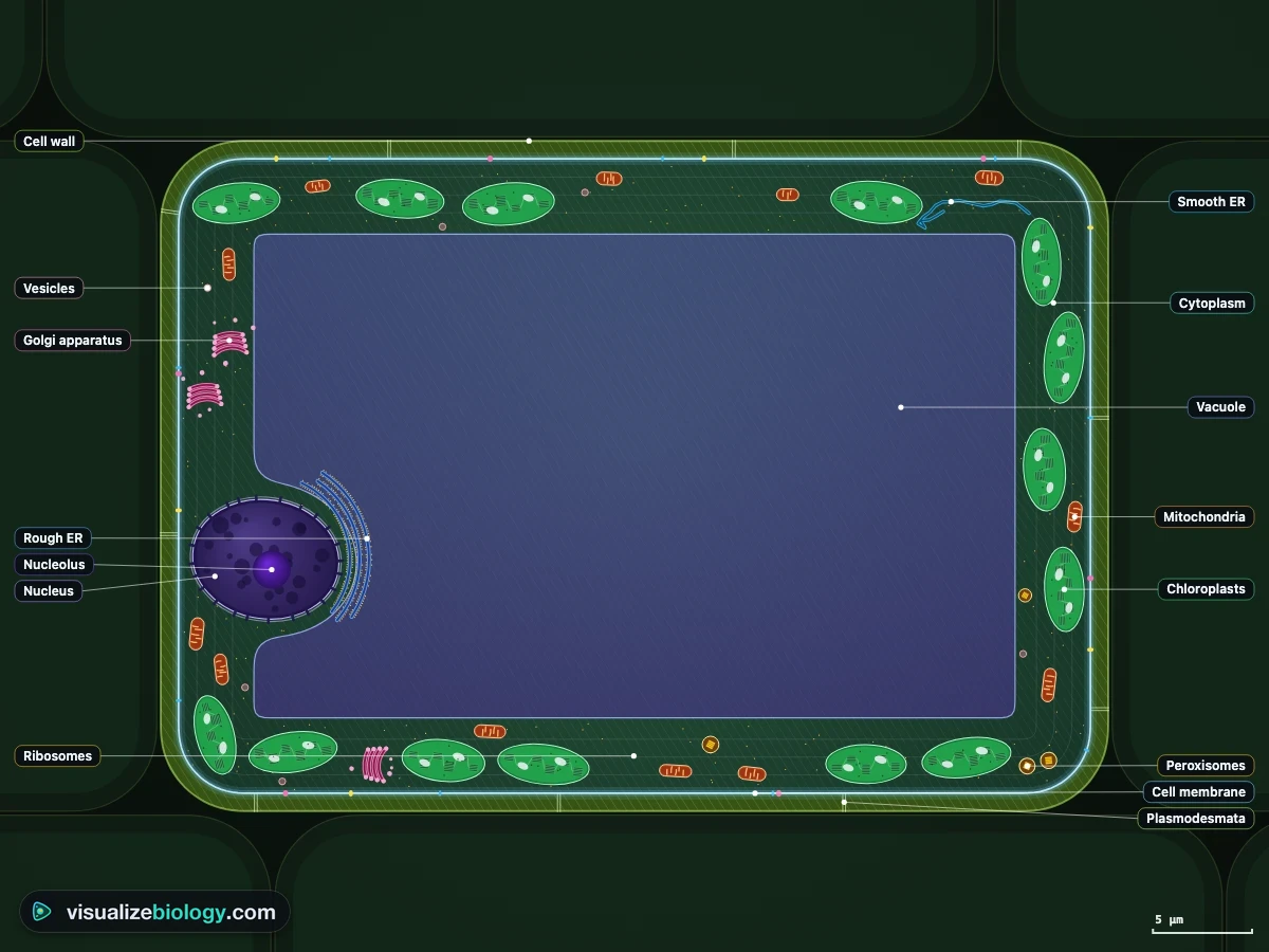
- Plant: a typical plant cell, a palisade leaf cell (packed with chloroplasts) and a root hair cell (a long hair for taking up water and minerals, no chloroplasts).
- Cells and organelles are drawn to scale with each other; the scale bar in the corner follows the zoom. Not every organelle is drawn: the Organelle panel says how many a real cell has.
3 Exploring the parts
- Hover over any part to see its name; tap it to select it. The Organelle panel gives its structure, membrane, size, how many there are and what it does, and the Close-up view draws it from inside, moving.
- Labels names every part with a line to it. Zoom with the + and - buttons, Ctrl or ⌘ + scroll, a double-click or a pinch, and press ⤢ to see the whole cell again. Once zoomed in, drag to look around.
- The Animal vs plant view lists every structure and which kind of cell has it; tap a row to find it in the cell.
4 A living cell: energy and the clock
The cell's clock runs in real time (or 10× or 60× faster, in the View panel). All the time, the cell uses ATP for its pumps, for making proteins and for everything else, and remakes it by respiration in its mitochondria. With Molecules on, glucose (yellow hexagons) and oxygen (red pairs) come in, carbon dioxide leaves and ATP (gold sparks) flies from the mitochondria to where it is used; one dot stands for millions of molecules. The Energy view shows where the energy goes, and Graph records ATP, oxygen and volume over time.
5 Surroundings, activity and poisons
- Surroundings: oxygen, glucose (or the sugar supply of a plant cell), temperature, light and carbon dioxide (plant cells) and the concentration of the outside solution.
- Activity: how hard the cell works. Cyanide stops the electron transport chain, ouabain stops the sodium-potassium pump (so the cell swells) and cycloheximide stops ribosomes making proteins.
- A cell that runs out of ATP for long enough, overheats or bursts dies; press ↺ New cell for a fresh one.
6 Show me: the guided animations
Each Show me button runs a short animation in steps, with the camera moving in on the parts that matter and the close-up following along: Respiration follows one glucose molecule from the blood to ATP; Photosynthesis follows light, water and carbon dioxide into a chloroplast and the sugar out of it; Make a protein follows the human insulin gene from DNA to mRNA, the ribosome, the rough ER, the Golgi apparatus and out of the cell; Osmosis puts the cell in water and in salt; Transport shows diffusion, carriers, pumps and vesicles; Engulf (animal cells) shows a bacterium eaten and digested by lysosomes. Use ‹ and › to step, ❚❚ to pause and Done to finish.
7 Experiment mode and the lab report
Seven labs: label the parts of animal and plant cells, measure a cell with the ruler and calculate the magnification, respiration with and without oxygen, temperature and respiration, light and photosynthesis in a leaf cell, osmosis in animal and plant cells, and from gene to protein with mutations. Each shows its aim, what you need and a real-lab safety note; the method ticks off as you work, a conclusion question checks your result, and Create lab report builds a PDF with your cell, results and calculations.
8 Quiz and units
The practice quiz covers organelles, animal and plant cells, size and magnification, respiration, photosynthesis, genes and proteins, osmosis and transport, and specialized cells, each with a worked solution. Take the quiz gives five mixed questions and a star rating. The SI (°C) | US (°F) switch changes temperatures; cell sizes stay in micrometers (µm), as in every biology class.
9 Keyboard and gestures
- Space pauses or runs the clock, F shows the whole cell, + and − zoom, S opens Status, Esc clears the selection or stops an animation, and ← and → step through an animation.
- A plain scroll over the cell scrolls the page; hold Ctrl or ⌘ (or click the cell first) to zoom with the wheel. On a phone, one finger scrolls the page and two fingers zoom and move the cell.
- Right-click the cell or the close-up to save it as an image or copy the readings.
Animal and plant cells: the parts and what they do
Every living thing is made of cells, and animals and plants are built from the same kind: the eukaryotic cell, with its DNA inside a nucleus and its work divided among membrane-bound organelles. This interactive cell model lets you look at both, part by part, and then watch them work. Choose a typical animal or plant cell, or one of five specialized cells, tap any organelle to see its structure, size and job, and open the close-up to see it from the inside.
- Cells
- Typical animal and plant cells, a liver cell, a pancreatic β-cell, a macrophage, a palisade leaf cell and a root hair cell
- Organelles
- Nucleus, nucleolus, ribosomes, rough and smooth ER, Golgi, mitochondria, chloroplasts, vacuole, lysosomes, peroxisomes, centrioles, cytoskeleton, membrane, wall
- It shows
- Respiration and ATP, photosynthesis, protein synthesis from the insulin gene, osmosis, membrane transport, phagocytosis
- Modes
- Simulate (with guided animations), Experiment (7 labs) and Quiz
- Price
- Free, with no account or download; works on Chromebooks, tablets and phones
Organelles and their functions
| Organelle | Structure | Function |
|---|---|---|
| Nucleus | Double membrane with pores; DNA as chromatin; nucleolus | Holds the genes and controls the cell |
| Ribosomes | rRNA and protein, 25–30 nm, free or on the rough ER | Make proteins (translation) |
| Rough ER | Membrane sheets studded with ribosomes | Folds and ships proteins for export |
| Smooth ER | Membrane tubules, no ribosomes | Makes lipids and steroids, breaks down toxins |
| Golgi apparatus | Stack of flattened sacs | Modifies, sorts and packages proteins into vesicles |
| Mitochondria | Double membrane, inner one folded into cristae | Aerobic respiration: make most of the ATP |
| Chloroplasts (plant) | Double membrane, thylakoids stacked in grana | Photosynthesis: make glucose |
| Vacuole | Plant: one large sac of sap, the tonoplast; animal: small | Stores sap, keeps a plant cell turgid |
| Lysosomes (animal) | Sacs of digestive enzymes at pH 4.8 | Digest bacteria and worn-out parts |
| Cell membrane | Phospholipid bilayer with proteins | Controls what enters and leaves |
| Cell wall (plant) | Cellulose fibers, fully permeable | Supports the cell, stops it bursting |
Animal cell vs plant cell
Animal and plant cells share the nucleus, cytoplasm, cell membrane, mitochondria, ribosomes, ER and Golgi apparatus. Three structures are found only in plant cells: the cellulose cell wall, chloroplasts and a large permanent vacuole. Animal cells have centrioles and more lysosomes, no wall and only small, temporary vacuoles, so they are rounder and more variable in shape; plant cells are usually larger (10–100 µm against 10–30 µm) and boxier. Animals store glucose as glycogen, plants as starch. Not every plant cell has chloroplasts: a root hair cell lives in the dark soil and has none.
A living cell: respiration and ATP
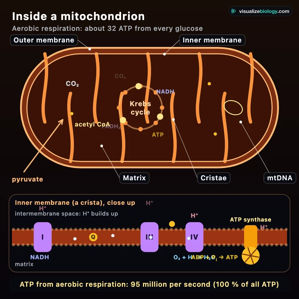
A cell is never still. A typical human cell uses about 100 million molecules of ATP every second, for its pumps, for making proteins and for building and moving, and its whole store of ATP would last only a minute or two if it were not remade. It is remade by respiration: glucose + oxygen → carbon dioxide + water, releasing energy. Glycolysis in the cytoplasm splits glucose into two pyruvate and gives 2 ATP; in the mitochondria the link reaction and the Krebs cycle release the carbon as CO₂ and load NADH, and the electron transport chain on the cristae uses oxygen to make about 28 more, about 32 in all. Without oxygen (or with cyanide, which blocks the chain), only glycolysis works: 2 ATP per glucose, with lactate in animal cells or ethanol and CO₂ in plants. Turn the oxygen off in the simulator and watch the cell burn glucose several times faster and still make less ATP.
Photosynthesis and the plant cell
In a leaf cell, chloroplasts capture light: on the thylakoid membranes water is split, releasing oxygen, and ATP and NADPH are made; in the stroma the Calvin cycle uses them to fix carbon dioxide into sugar (6 CO₂ + 6 H₂O → C₆H₁₂O₆ + 6 O₂). The glucose is used in respiration, stored as starch grains or sent to the rest of the plant as sucrose. The cell still respires day and night, so in dim light it takes in oxygen; above its compensation point it gives oxygen out. A palisade cell even moves its chloroplasts: they spread out in dim light and line up along the side walls in very bright light.
From gene to protein
Proteins are made in two steps. In transcription, RNA polymerase copies a gene's DNA into messenger RNA in the nucleus; the introns are spliced out and the mRNA leaves through a nuclear pore. In translation, a ribosome reads the mRNA one codon (three bases) at a time while transfer RNAs bring the matching amino acids. The simulator uses the real coding sequence of the human insulin gene: its first codons send the new protein into the rough ER, where it folds, then it passes through the Golgi apparatus into secretory granules, is cut into insulin and leaves the cell by exocytosis. Change one base and see whether the mutation is silent, missense, nonsense or a frameshift.
Water and the membrane: osmosis
The cell membrane is partially permeable: oxygen, carbon dioxide and water cross it freely, glucose needs a carrier and ions are moved by pumps that use ATP. Water moves by osmosis towards the more concentrated solution. In pure water an animal cell swells and bursts; in strong salt it shrinks and crenates. A plant cell in water swells only until its wall pushes back, becoming firm or turgid; in a solution stronger than its sap the protoplast pulls away from the wall, which is plasmolysis. That is why plants wilt when they lose water.
Experiments and course links
| Lab | What you find out | Course links |
|---|---|---|
| Label animal and plant cells | The parts of each and the three plant-only structures | NGSS MS-LS1-2; GCSE 4.1.1.2; NCERT Class 9 |
| Measure a cell | Image size, actual size and magnification | GCSE 4.1.1.5; AP Biology 2.3 |
| Respiration with and without oxygen | ATP per glucose and the Pasteur effect | HS-LS1-7; AP Biology 3.6 |
| Temperature and respiration | The optimum temperature and denaturing enzymes | HS-LS1-7; GCSE 4.4.2 |
| Light and photosynthesis | The light compensation point of a leaf cell | HS-LS1-5; AP Biology 3.5 |
| Osmosis in animal and plant cells | Bursting, crenation, turgor and plasmolysis | GCSE 4.1.3.2; AP Biology 2.8 |
| From gene to protein | Transcription, translation and mutations | HS-LS1-1; AP Biology 6.3–6.4 |
Who uses this cell model?
Students use it to learn the parts of animal and plant cells and to see what each one does, to revise respiration, photosynthesis and protein synthesis with moving pictures instead of static diagrams, and to practice magnification and osmosis questions with worked answers. Teachers project the guided animations, ask the class to predict what cyanide or pure water will do before trying it, and set the labs as homework. It is an educational model built from published cell biology; it needs no login and runs on school Chromebooks.
Frequently asked questions
What are the main differences between an animal cell and a plant cell?
Both are eukaryotic cells with a nucleus, cytoplasm, a cell membrane, mitochondria, ribosomes, endoplasmic reticulum and a Golgi apparatus. Plant cells also have a cellulose cell wall, chloroplasts for photosynthesis and a large permanent vacuole filled with cell sap, and they are usually larger and more regular in shape. Animal cells have no wall, no chloroplasts and only small temporary vacuoles, but they have centrioles and more lysosomes. Animal cells store glucose as glycogen; plant cells store it as starch.
What does each organelle do?
The nucleus holds the DNA and controls the cell; the nucleolus makes ribosomes. Ribosomes make proteins, the rough ER folds and ships proteins, the smooth ER makes lipids, and the Golgi apparatus modifies and packages them into vesicles. Mitochondria release energy from glucose by aerobic respiration, making most of the cell's ATP. Chloroplasts make glucose by photosynthesis. Lysosomes digest bacteria and worn-out parts, the vacuole stores sap and keeps a plant cell turgid, the cell membrane controls what goes in and out, and the cell wall supports a plant cell and stops it bursting.
How much ATP does a cell make?
A typical human cell uses and remakes roughly 100 million ATP molecules every second, so its whole store of ATP lasts only a minute or two. With oxygen, aerobic respiration releases about 30 to 32 ATP from each glucose molecule (older textbooks say 36 to 38): 2 from glycolysis in the cytoplasm, 2 from the Krebs cycle and about 28 from the electron transport chain in the mitochondria. Without oxygen, only glycolysis works, giving 2 ATP per glucose, with lactate (animals) or ethanol and carbon dioxide (plants and yeast) as the waste.
Explore related simulators
Decode any gene codon by codon, look up every codon on a codon wheel and see what a point mutation does in the Interactive Codon Chart and DNA to Protein Translator. Watch a cell divide in the Mitosis Simulator, and see how gametes are made in the Meiosis Simulator. Turn the molecule inside the nucleus in the DNA Structure 3D Model, measure what light and carbon dioxide do to a leaf in the Photosynthesis Lab, run potato and red blood cell osmosis practicals in the Osmosis Lab, see real onion and cheek cells in the Virtual Microscope, and meet a single cell that is a whole organism in the Virtual Amoeba Simulator. Compare these eukaryotic cells with a prokaryote in the Virtual Bacteria Simulator: no nucleus, a cell wall of peptidoglycan and 70S ribosomes. See how plant cells are joined into tubes that carry water and sugar through a whole plant in the Xylem and Phloem simulator. See where a cell's glucose and amino acids come from in the Human Digestive System Simulator, where villi absorb them, and where the oxygen for aerobic respiration comes from in the Respiratory System Simulator, which also sets breathing apart from cellular respiration.
Sources: OpenStax Biology 2e, chapters 4 (Cell Structure), 5 (Plasma Membranes), 7 (Cellular Respiration), 8 (Photosynthesis) and 15 (Genes and Proteins); Urry et al., Campbell Biology 12th edition, chapters 6–10 and 17; Alberts et al., Molecular Biology of the Cell 6th edition; Milo and Phillips, Cell Biology by the Numbers (2015); Nobel, Physicochemical and Environmental Plant Physiology; Bell et al., Nature 284 (1980), the human insulin gene (GenBank NM_000207); College Board, AP Biology Course and Exam Description. Sizes, rates and concentrations are an educational model based on these sources; nothing here is medical advice.
Updated