
From the microscope to the organelle
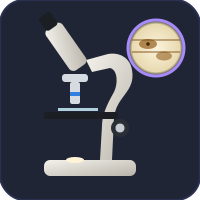
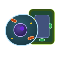
A cell biology unit usually begins at the microscope and ends inside the cell. The Virtual Microscope teaches focusing, magnification and measuring cells on real slides. The Virtual Living Cell then opens an animal and a plant cell whose organelles are working: mitochondria making ATP, ribosomes building insulin, a vacuole pushing on the wall.
Which simulation for which topic
| Topic | Open |
|---|---|
| Cell structure, organelles, magnification | Virtual Living Cell, Virtual Microscope |
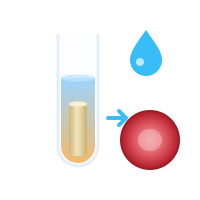
| Diffusion, osmosis and water potential | Osmosis Lab |
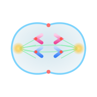
| The cell cycle and mitosis | Mitosis Simulator |
| Meiosis and gametes | Meiosis Simulator |
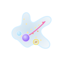
| A single-celled organism | Virtual Amoeba Simulator |
Why simulate a cell?
Most of what a cell does is too small, too fast or too slow to see in a school lab. A dividing onion cell takes hours; water crossing a membrane cannot be seen at all. In a simulation you can slow time down, label every structure and change one condition at a time, such as the sugar concentration around a potato core or the drug added to a dividing cell, then read the result off a graph. Each page also has the practical you would do in a real lab, with a results table and a lab report.
Updated