
Mitosis Simulator
Watch a cell copy its chromosomes and divide in 3D, stage by stage. Break its checkpoints to see how cancer starts, then count real-looking onion root tip cells to find the mitotic index, as in AP Biology Lab 7.
Live math
Lab notebook
Every count you record, and every event in the cell (a checkpoint holding, nondisjunction, apoptosis), is written here with its conditions. Export a CSV, or turn the session into a PDF lab report.
| # | Sample | Where / when | I · P · M · A · T | Total | Result |
|---|
Concept library
Click "New problem" to begin.
Click "Start quiz" for 5 questions: naming the stage, chromosome counts, mitotic index math, cell cycle control and lab skills.
1 Overview
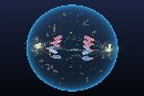
Simulate shows one cell going round the whole cell cycle: interphase (G1, S, G2), then prophase, prometaphase, metaphase, anaphase, telophase and cytokinesis. Choose an animal or a plant cell, play it, step through it phase by phase, and look at it from any side in 3D. Beside it you can watch the cell cycle wheel with its checkpoints, a close-up of one pair of chromosomes, graphs of DNA content, chromosome number and cyclins, and a root tip slide. Experiment is a virtual onion root tip lab with six labs to carry out yourself, from the squash to a chi-square test, ending in a PDF lab report. Quiz has a practice quiz with worked solutions and a 5-question quiz.
2 Getting started
- The cell opens in 3D on a computer or tablet and in 2D on a phone. The 2D | 3D switch changes it any time and remembers your choice.
- Press ▶ (or Space) to play the cycle. Teach speed gives every phase a few seconds on screen; the other speeds run in the cell's real time, so you see how much longer interphase is than mitosis.
- Next phase ⏭ (or N) jumps to the start of the next phase and pauses, so you can study each one; or switch on Pause at each new phase and press ▶ after each. Click any segment of the phase strip, the cycle wheel or the graph to jump straight there.
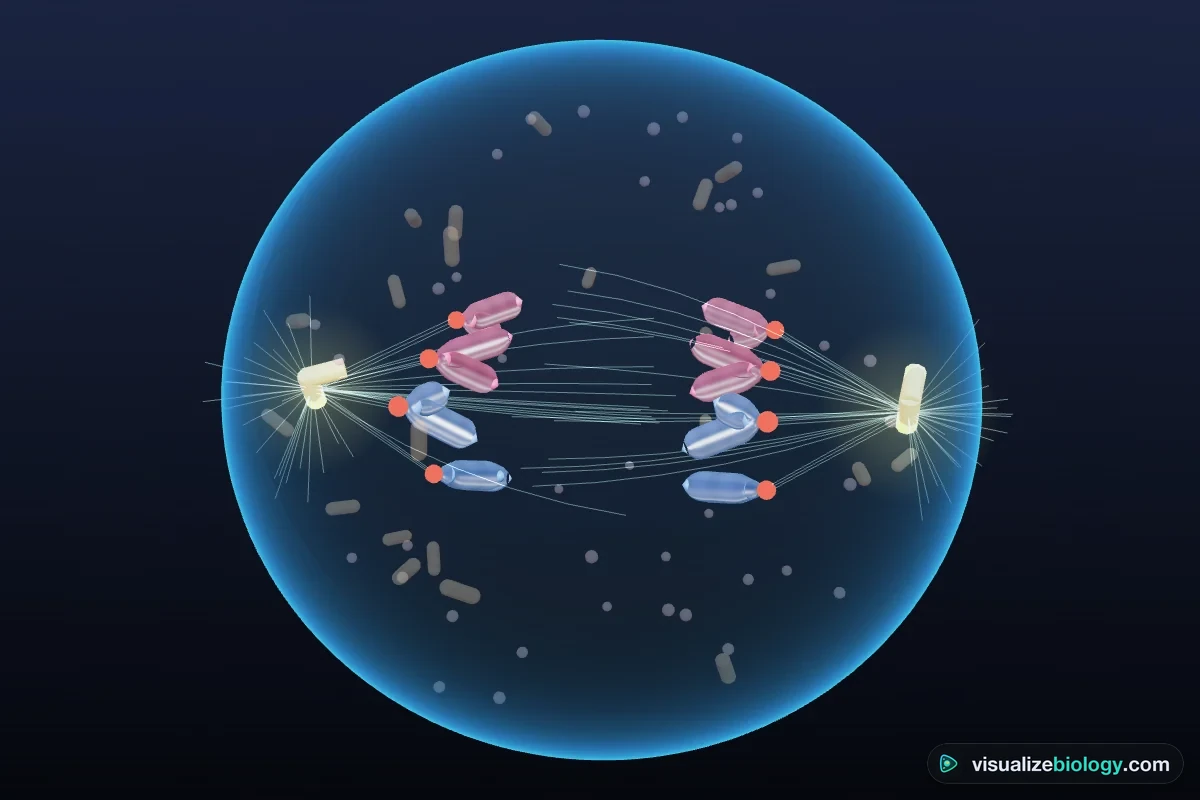
- The caption on the cell says what is happening at that moment, and the arrows show where things are moving: centrosomes, chromosomes on their way to the plate, chromatids to the poles, the furrow pinching in or the cell plate growing.
- Click a part of the cell (a chromosome, the spindle, a centrosome) to name it and read what it does.
3 The cell (Simulate)
- Animal or plant: the animal cell has centrosomes with centrioles and asters, and divides with a cleavage furrow. The plant cell (an onion root tip cell) has a cell wall, no centrioles, a preprophase band, and divides with a cell plate.
- Chromosomes: 2n = 4, 6 or 8 keeps the picture clear; the last button shows the real number (46 for a human cell, 16 for onion). Chromosomes from the mother are pink and from the father blue, so you can follow the homologous pairs. Follow one chromosome pair fades all the others, so you can watch one maternal and one paternal chromosome copy, line up, split, and end up once in each daughter cell.
- Fluorescent draws the cell as a cell biologist sees it in a fluorescence microscope: DNA blue, microtubules green, kinetochores red.
- Conditions: temperature changes the length of the cycle (Q₁₀ = 2); too cold or too hot and the cell stops. Without growth factors the cell leaves the cycle into G0. Colchicine stops the spindle forming, so the cell is held in metaphase.
4 Checkpoints and cancer
- ☢ Damage DNA breaks a chromosome. With the G1 or G2 checkpoint on, the cell stops until the break is repaired; three breaks and p53 sends it into apoptosis. With the checkpoints off, the damage is copied into the daughter cells as a mutation (a yellow mark).
- ✂ Detach a kinetochore leaves one chromatid unattached. The spindle checkpoint holds anaphase until it is fixed. Turn the checkpoint off and both chromatids go to the same pole: nondisjunction. The daughter you follow then has an extra chromosome.
- 🦠 Cancer cell switches on an oncogene (the cell divides without growth factors) and removes p53 (the G1 and G2 checkpoints).
5 Cycle, Chromosomes, Graph and Slide views
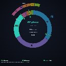
- Cycle: the wheel in real proportions, with mitosis magnified on an outer ring, the three checkpoints (red when holding) and G0. The middle shows the chromosome number, sister chromatids and DNA content.
- Chromosomes: one homologous pair close up: chromatin, then two sister chromatids, centromere, kinetochores and alleles of a gene. The A-level / AP chip adds cohesin.
- Graph: DNA per cell (2C to 4C and back), chromosomes per cell, or cyclins and CDK, with a cursor that follows the cell.
- Slide: an onion root tip squash. In Simulate, Show every cell's stage outlines each dividing cell in its stage's color, and Auto-count counts the field for you.
6 The onion root tip lab
Prepare the squash with the buttons on the bench (cut, hydrolyze in HCl, rinse, stain, squash), or skip a step to see what goes wrong. Then drag the slide to move it, and use 100× to find the zone of cell division and 400× to count. Click a cell and choose its stage, or hover over it and press I, P, M, A or T. Count every cell in a field so you do not pick only the interesting ones. Your tally, the mitotic index and the bar chart update as you go. The cell on the left shows the stage you just chose, in 2D or 3D.
7 Experiment mode and the lab report
Six labs: the mitotic index (AP Lab 7, A-level Required Practical 2), how long each stage lasts, where in the root mitosis happens, lectin and the chi-square test (AP Lab 7 Part 2), colchicine over time, and the whitefish blastula. Each shows its aim, what you need and a real-lab safety note. The method ticks off as you do each step, a conclusion question checks the result from your own counts, and Create lab report builds a PDF with your tally, graph, worked calculations and sources of error.
8 Quiz
The practice quiz asks you to name the stage from a drawing, count chromosomes, chromatids and DNA, calculate mitotic indexes, stage times and chi-square, and explain checkpoints, cancer and the lab method, with a worked solution for each. Take the quiz gives five mixed questions and a star rating.
9 Keyboard and gestures
- Space plays or pauses, N goes to the next phase, S opens Status, and C, H, G switch to Cycle, cHromosomes or Graph.
- On a slide, hover over a cell and press I, P, M, A or T to count it; Z undoes the last one.
- Scrolling over the 3D cell always scrolls the page. To zoom, hold Ctrl (⌘ on a Mac) and scroll, or pinch on a trackpad. On a phone, one finger scrolls the page; turn the 3D cell with two fingers.
- Right-click a view to save it as an image or copy your results.
Stages of mitosis: an interactive mitosis simulator and onion root tip lab
Mitosis is how one cell divides into two genetically identical daughter cells. It is how you grew from a single fertilized egg, how a cut heals and how a plant's root pushes through the soil. This free mitosis simulator lets you watch it happen in 3D: the chromosomes condense, line up, split and are shared out, and the cell pinches in two. You can slow it down, step through it one phase at a time, and break the controls that keep it accurate to see what goes wrong in cancer. Then, in the virtual lab, you count cells on an onion root tip slide to measure mitosis yourself, as in AP Biology Lab 7.
- Cells
- An animal cell (human, 2n = 46) and a plant cell (onion root tip, 2n = 16), or 2n = 4, 6 or 8 for clarity
- Phases
- G1, S, G2, prophase, prometaphase, metaphase, anaphase, telophase and cytokinesis
- Regulation
- G1, G2 and spindle checkpoints, cyclins and CDK, growth factors and G0, p53 and apoptosis, nondisjunction, colchicine, a cancer cell
- Slides
- Onion root tip squash (100× and 400×), lectin-treated and colchicine-treated root tips, whitefish blastula
- Measurements
- Mitotic index, time in each stage, mitotic index along the root, chi-square test
- Modes
- Simulate, Experiment (6 labs) and Quiz
- Runs on
- Any modern browser, including Chromebooks, iPads and phones. 3D on larger screens, 2D on phones.
- Price
- Free, with no account or download
What is mitosis?
Mitosis is the division of the nucleus that gives two nuclei with exactly the same chromosomes as the parent nucleus. It is followed by cytokinesis, the division of the cytoplasm, so one diploid cell becomes two identical diploid cells. A human cell starts with 46 chromosomes and each daughter cell ends with 46. Mitosis is used for growth, for repairing and replacing cells, and for asexual reproduction. It is different from meiosis, which makes gametes with half the number of chromosomes.
Before a cell can divide it must copy its DNA. That happens in interphase, the long part of the cell cycle between divisions: the cell grows in G1, copies every chromosome in S phase (so each one becomes two identical sister chromatids), and grows and checks its DNA in G2. In the onion root tip cells in this simulator, interphase takes about 17.8 of the 20 hours of the cycle; mitosis takes only about 2.2 hours.
The phases of mitosis in order (PMAT)
The stages of mitosis are prophase, metaphase, anaphase and telophase, remembered as PMAT. AP Biology and most college textbooks add prometaphase between prophase and metaphase. Cytokinesis usually begins during late anaphase or telophase.
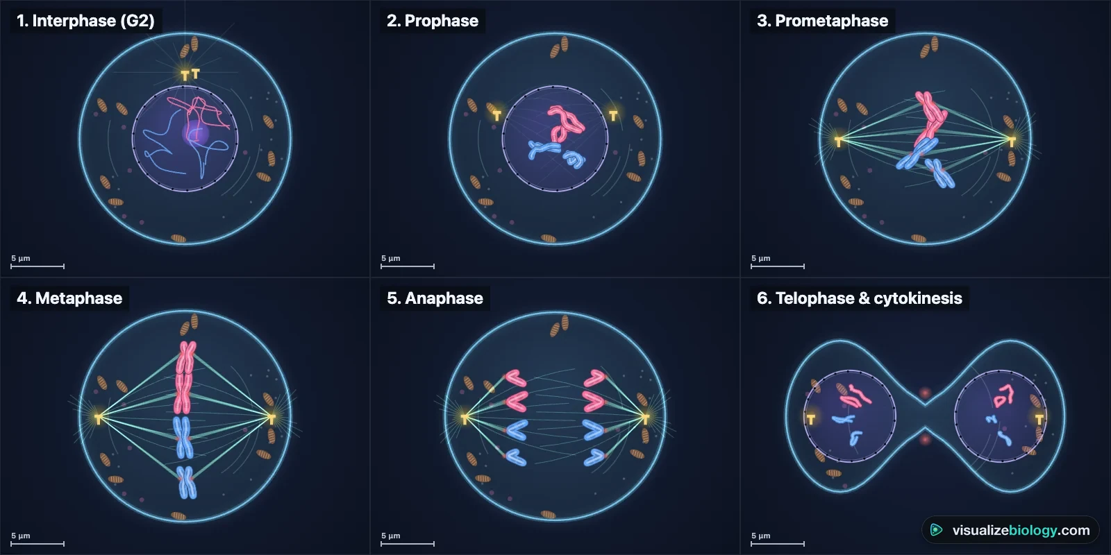
| Stage | What happens | How to spot it under the microscope |
|---|---|---|
| Prophase | Chromatin condenses into chromosomes, each two sister chromatids joined at a centromere. The nucleolus disappears. The centrosomes move apart and the spindle begins to form. | A round nucleus full of thick, tangled threads |
| Prometaphase | The nuclear envelope breaks into fragments. Spindle microtubules attach to the kinetochore on each sister chromatid. | Condensed chromosomes loose in the cytoplasm, no nuclear envelope |
| Metaphase | Chromosomes line up on the metaphase plate at the cell's equator, each attached to both poles. | A dark band of chromosomes across the middle of the cell |
| Anaphase | Cohesin is cut, the sister chromatids separate and are pulled to opposite poles (anaphase A) while the poles move apart (anaphase B). | Two V-shaped groups moving apart, arms trailing behind |
| Telophase | A nuclear envelope re-forms round each set of chromosomes, which uncoil. The spindle breaks down. | Two dark groups at opposite ends, often with a new cell wall between |
| Cytokinesis | The cytoplasm divides: a cleavage furrow in animal cells, a cell plate in plant cells. | Two daughter cells |
Chromosomes, chromatids and DNA during the cell cycle
Counting gets confusing because the words change meaning. A chromosome is counted by its centromere. After S phase each chromosome is two sister chromatids, but it is still one chromosome. In anaphase the sister chromatids separate, and each one is now counted as a chromosome, so for a moment a human cell holds 92 chromosomes. The DNA content doubles from 2C to 4C during S phase and halves at cytokinesis. The Chromosomes view and the Graph view in the simulator show these numbers at every moment.
| Human cell | Chromosomes | Sister chromatids | DNA content |
|---|---|---|---|
| G1 | 46 | 0 | 2C |
| G2, prophase, metaphase | 46 | 92 | 4C |
| Anaphase | 92 | 0 | 4C |
| Each daughter cell | 46 | 0 | 2C |
Mitosis in plant and animal cells
The stages are the same, but plant cells do it without centrioles and inside a rigid wall. An animal cell organizes its spindle from two centrosomes, each with a pair of centrioles and a star-shaped aster of microtubules, and divides by a cleavage furrow: a ring of actin and myosin under the membrane contracts and pinches the cell in two. A plant cell has no centrioles; its spindle forms from the nuclear envelope and wide poles, a preprophase band of microtubules marks where the new wall will be, and in telophase vesicles carrying wall material gather on a framework of microtubules (the phragmoplast) and fuse into a cell plate that grows outward until it meets the old walls. Switch between the animal and plant cell in the simulator to compare them side by side.
Cell cycle checkpoints, cyclins and cancer
The cell cycle is driven by cyclins and cyclin-dependent kinases (CDKs). Each cyclin rises at its own point in the cycle and switches on its CDK; cyclin B with CDK1 (MPF) starts mitosis, and its destruction at anaphase lets the cell finish. Three checkpoints stop the cycle when something is wrong. The G1 checkpoint checks for growth signals and DNA damage, and without growth factors the cell rests in G0. The G2 checkpoint checks that DNA has been copied without damage. The spindle assembly checkpoint holds anaphase until every chromosome is attached to both poles. The protein p53 holds a damaged cell in G1 and, if the damage is too great, starts apoptosis.
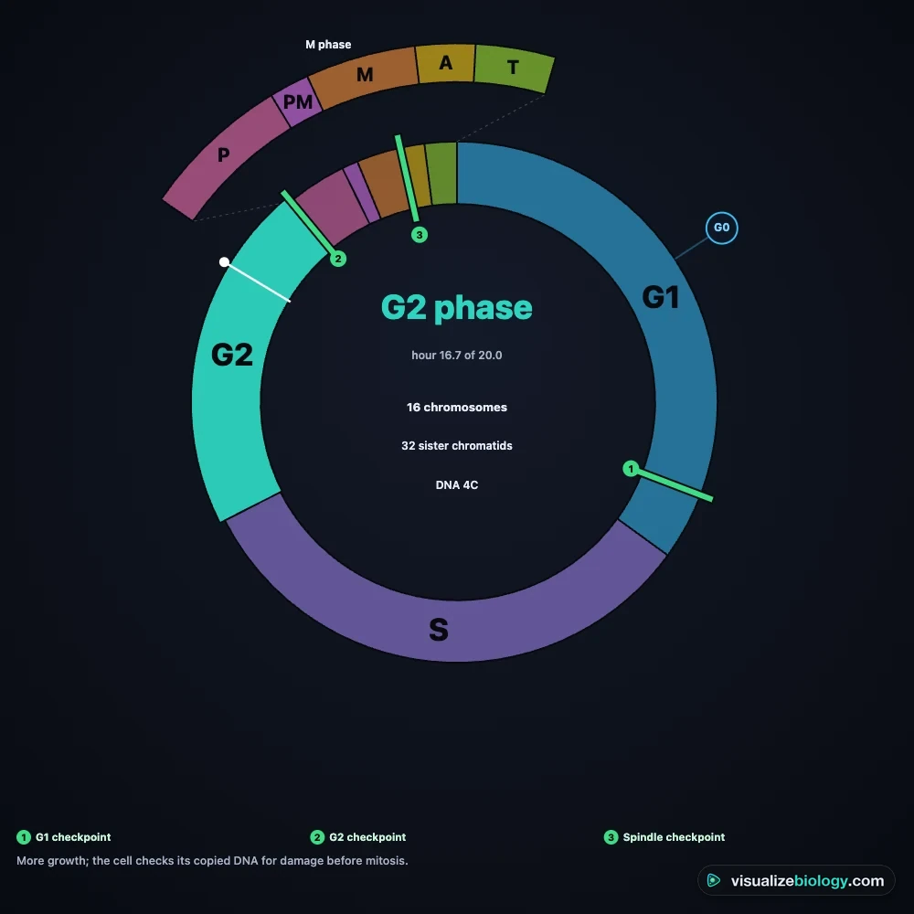
Cancer is cell division out of control. Mutations in proto-oncogenes make a cell divide without growth signals, and mutations in tumor suppressor genes such as p53 remove the checkpoints, so damaged DNA is copied and chromosomes are shared out unequally. In the simulator, switch off the spindle checkpoint and detach a kinetochore to see nondisjunction: both chromatids go to the same pole and the daughter cell gets an extra chromosome. Many chemotherapy drugs, such as taxol and the vinca alkaloids, attack the spindle as colchicine does.
The onion root tip lab and the mitotic index
The growing tip of a root is the classic place to see mitosis. Behind the root cap is the meristem, the zone of cell division, where new cells are made; behind that, in the zone of elongation, cells stop dividing and grow longer. Root tips are hydrolyzed in hydrochloric acid, stained so the chromosomes show up, and squashed into a single layer of cells. Under the microscope, you classify and count the cells.
mitotic index = cells in mitosis ÷ total cells counted × 100
time in a stage = (cells in that stage ÷ total cells counted) × length of the cell cycle
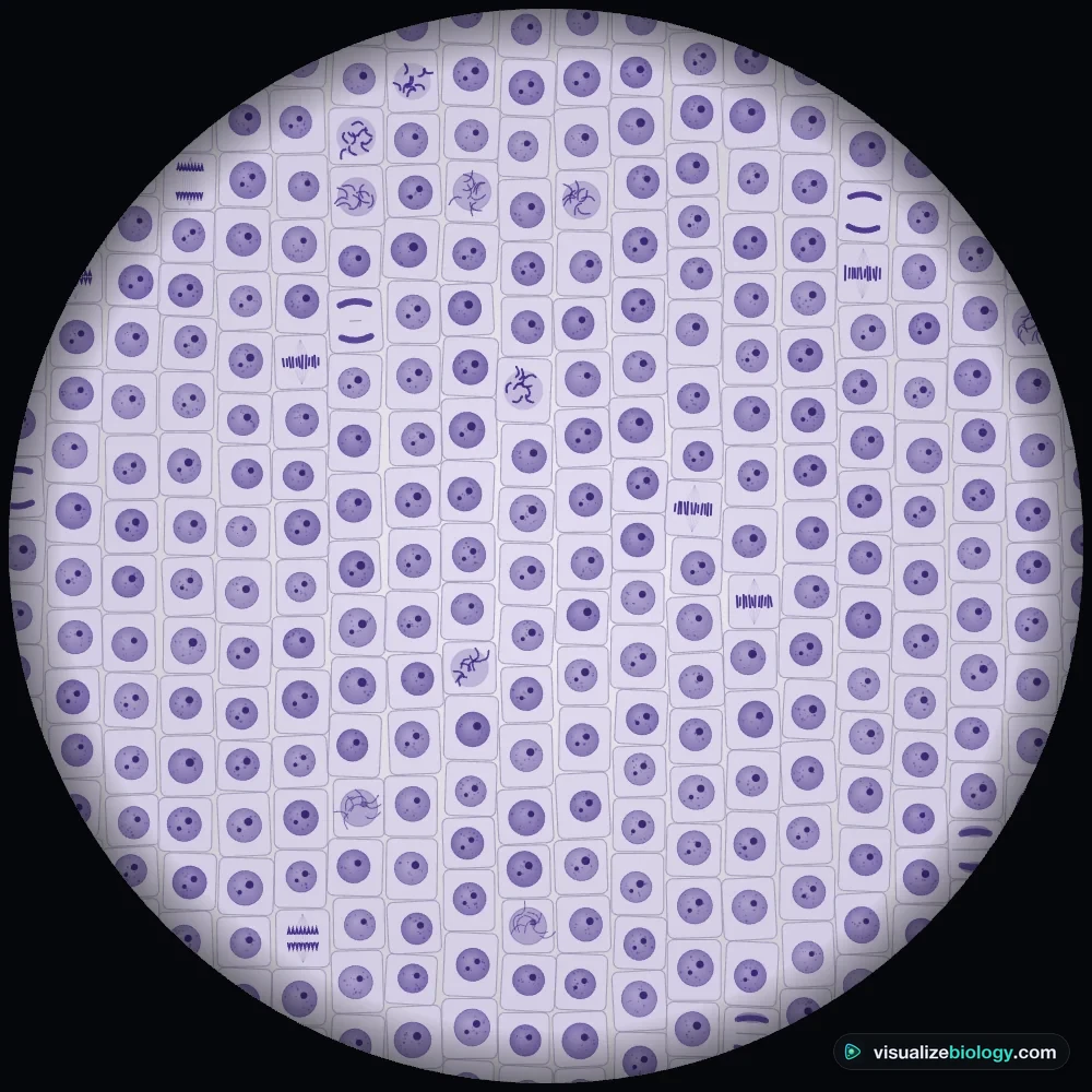
Worked example. A student counts 200 cells in the meristem: 178 in interphase, 10 in prophase, 5 in metaphase, 3 in anaphase and 4 in telophase. The mitotic index is 22 ÷ 200 × 100 = 11 %. If the cell cycle lasts 20 hours, metaphase takes 5 ÷ 200 × 20 h = 0.5 h, or 30 minutes. Prophase has the most cells, so it is the longest stage of mitosis; anaphase has the fewest, so it is the shortest. This works because the cells are dividing out of step with each other, so the fraction of cells in a stage matches the fraction of time spent in it.
AP Biology Lab 7: lectin and the chi-square test
In Part 2 of AP Biology Investigation 7, root tips are grown with and without lectin, a protein that can trigger cell division, and the numbers of cells in interphase and in mitosis are compared with a chi-square test. The null hypothesis is that lectin has no effect. Expected values come from the totals: expected = row total × column total ÷ grand total. With 1 degree of freedom the critical value at p = 0.05 is 3.84.
| Root tips | Interphase | Mitosis | Total |
|---|---|---|---|
| Control | 178 (expected 171) | 22 (expected 29) | 200 |
| Lectin | 164 (expected 171) | 36 (expected 29) | 200 |
χ² = 7²/171 + 7²/29 + 7²/171 + 7²/29 = 0.29 + 1.69 + 0.29 + 1.69 = 3.95, which is greater than 3.84, so the null hypothesis is rejected: lectin increased the proportion of cells in mitosis. In the simulator your own counts give your own chi-square, and with a small sample it may not reach significance, as in a real lab.
Experiments and course links
| Lab | What you find out | Course links |
|---|---|---|
| Onion root tip: mitotic index | Prepare a squash and calculate the mitotic index | AP Biology Lab 7, NGSS HS-LS1-4, A-level RP2 |
| How long is each stage? | Stage times from cell counts and a 20-hour cycle | AP Topic 4.6, A-level, IB D2.1 |
| Where does mitosis happen? | The mitotic index falls from the meristem to the zone of elongation | NGSS HS-LS1-4, GCSE, IGCSE |
| Lectin and chi-square | Does lectin increase cell division? A χ² test of independence | AP Biology Lab 7 Part 2 |
| Colchicine over time | Cells pile up in metaphase when the spindle cannot form | AP Topic 4.7, A-level |
| Whitefish blastula | Mitosis in fast-dividing animal cells, with asters | AP Biology, college intro biology |
Mitosis is one of two ways a nucleus divides. The other, meiosis, makes sperm and eggs with half the chromosomes, and its first division pairs up the homologs and shuffles their genes. For a stage-by-stage comparison of mitosis vs meiosis, see the Meiosis Simulator.
Who uses this mitosis simulator?
Students use it to learn the phases of mitosis in order, to see why chromosome numbers change, and to practice mitotic index, stage time and chi-square calculations with worked answers. Teachers project the 3D cell, pause it on each stage, and assign the onion root tip lab when there are no fresh roots or not enough microscopes. It is an educational model built from textbook values, it needs no login, and it runs on school Chromebooks.
Frequently asked questions
What are the stages of mitosis in order?
Prophase, metaphase, anaphase and telophase (PMAT), followed by cytokinesis. Many courses add prometaphase between prophase and metaphase, when the nuclear envelope breaks down and the spindle captures the chromosomes. Interphase (G1, S and G2) comes before mitosis but is not a stage of it.
What is the difference between a chromosome and a chromatid?
A chromosome is one DNA molecule with its proteins, counted by its centromere. After DNA is copied in S phase, each chromosome is made of two identical sister chromatids joined at the centromere. In anaphase the sister chromatids separate, and from then on each one is counted as a chromosome. A human cell has 46 chromosomes and, from S phase to metaphase, 92 chromatids.
How do you calculate the mitotic index?
Mitotic index = (number of cells in mitosis ÷ total number of cells counted) × 100. Count every cell in a field of view at 400x, including those in interphase. For example, 22 dividing cells out of 200 counted gives a mitotic index of 11 %.
Why are onion root tips used to study mitosis?
Just behind the root cap is the meristem, the zone of cell division, where many cells are dividing at any moment. Onion cells have only 16 chromosomes, which are large and easy to see, and root tips are cheap and quick to grow in water. Hydrochloric acid softens the tissue so it squashes into a single layer of cells.
How is cytokinesis different in plant and animal cells?
In an animal cell a ring of actin and myosin contracts under the membrane, making a cleavage furrow that pinches the cell in two. A plant cell has a rigid wall, so vesicles from the Golgi gather at the equator and fuse into a cell plate that grows outward until it joins the side walls and becomes a new wall.
What happens if a cell cycle checkpoint fails?
Checkpoints stop the cycle until a problem is fixed: the G1 and G2 checkpoints hold a cell with damaged DNA (or trigger apoptosis through p53), and the spindle checkpoint holds anaphase until every chromosome is attached to both poles. If a checkpoint fails, damaged DNA is copied into daughter cells and chromosomes can be shared unequally (nondisjunction). These errors build up in cancer cells.
Explore related simulators
Explore every organelle of the cell that divides, and watch it make ATP and proteins, in the Virtual Living Cell. Look at real cells and their nuclei with the Virtual Microscope, see how water moves into root cells in the Osmosis Lab, find out how plants make the sugar that fuels all this growth in the Photosynthesis Lab, and follow the other kind of cell division, the one that makes gametes, in the Meiosis Simulator. The DNA copied in S phase is shown atom by atom in the DNA structure 3D model. A single-celled amoeba also divides by mitosis, in binary fission: watch it in the Virtual Amoeba Simulator.
Sources: OpenStax Biology 2e, chapter 10 Cell Reproduction; Alberts et al., Molecular Biology of the Cell, 6th ed., chapter 17 The Cell Cycle; Van't Hof, Experimental Cell Research 51 (1968), on the cell cycle of root meristems; College Board, AP Biology Investigative Labs, Investigation 7. The cycle lengths and stage durations are an educational model based on these sources (onion root tip about 20 h at 20 °C, human cell in culture about 24 h at 37 °C).
Updated