
Virtual Amoeba Simulator
A living Amoeba proteus you can watch in 3D: it crawls on pseudopods, hunts and digests prey, pumps out water, breathes through its membrane and splits in two. Change its world, break it on purpose, then run seven virtual amoeba labs.
Live math
Lab notebook
Every measurement you record, and every event in the amoeba's life (a meal, a division, a burst), is written here with its conditions. Export a CSV, or turn the session into a PDF lab report.
| # | Lab / sample | Conditions | Measurement | Result |
|---|
Concept library
Click "New problem" to begin.
Click "Start quiz" for 5 questions: the parts of an amoeba, movement, feeding, water balance, respiration and reproduction.
1 Overview
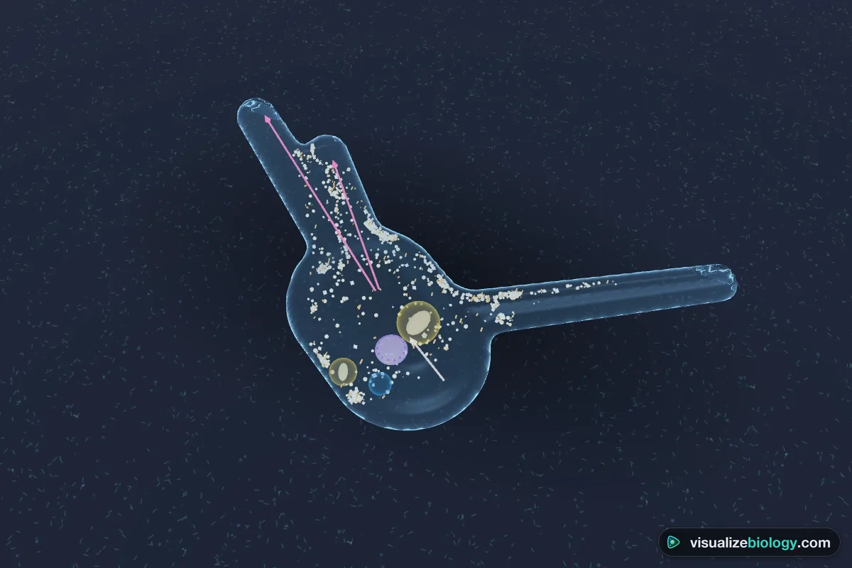
Simulate shows a living Amoeba proteus on a microscope slide. It crawls, hunts and eats prey, pumps out water, breathes and, when it has grown enough, divides in two. Change its water, temperature, oxygen and food, use the tools on the slide (needle, light, salt, scalpel), and break it on purpose. Beside it, Inside shows what you cannot see down a microscope (how a pseudopod works, a food vacuole being digested, osmosis and the contractile vacuole, gas exchange, the life cycle), and Graph plots the model. Experiment has seven labs that end in a PDF lab report, and Quiz has a practice quiz with worked solutions and a 5-question quiz.
2 Getting started
- The amoeba opens in 3D on a computer or tablet and in 2D on a phone. The 2D | 3D switch changes it and remembers your choice. Textbook | Microscope changes the look: colored diagram, or grey and granular as it really looks at 100×.
- Press ▶ (or Space). Teach runs 10× faster than real life (an amoeba is slow) and paces division so each stage gets a few seconds; the other speeds go from real time to one hour per second.
- Use Show in the top bar to jump to a process: movement, feeding, water balance, breathing, binary fission or cysts.
- Click any part of the amoeba to name it and read what it does. The caption says what is happening now, and the arrows show the cytoplasm streaming into the growing pseudopod.
- Open Status (S) for live readings: speed, length, volume, contractile vacuole rate, oxygen, surface area to volume ratio and doubling time.
3 Tools on the slide
- 👆 Look: click a part to name it; click another amoeba to follow it.
- 📏 Measure: drag a line to measure in µm.
- 🦠 Prey: click to drop prey (choose which in the Food panel). Amoebae find prey by chemotaxis and catch it by phagocytosis.
- 🪡 Needle: touch the amoeba; it pulls back the pseudopods there and turns away.
- 💡 Light and 🧂 Salt: Amoeba proteus avoids bright light and salt: watch a pseudopod that runs into them retract.
- 🔪 Cut: drag a glass needle across the amoeba to cut it in two. Only the half with the nucleus goes on feeding, growing and dividing.
- 🏷️ Label is for the labeling lab: click a part and choose its name.
4 Water, food and breaking it
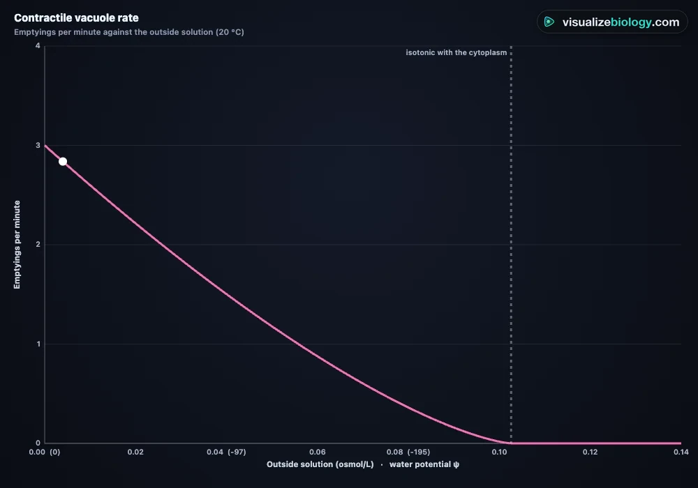
- Water: distilled, pond, brackish or seawater, plus sucrose. In fresh water the contractile vacuole fills and empties a few times a minute; as the outside solution approaches the cytoplasm (about 0.10 osmol/L) it slows and stops; in stronger solutions the amoeba shrinks and its surface crinkles. Temperature changes speed and growth (Q₁₀ ≈ 2; too hot, above about 33 °C, it dies). Oxygen controls how much ATP it can make: without it the amoeba slows and stops.
- Food: how much prey swims around, prey you drop, and neutral red stain to see a food vacuole turn acid.
- Break it: block the contractile vacuole (it swells and bursts in about an hour in pond water), harsh conditions (it rounds up and forms a cyst, then divides inside it), divide now, a light or a salt crystal in its path, cut it in two.
5 Inside and Graph
- Inside · Move: the sol–gel cycle in a pseudopod, at the amoeba's real speed. Feed: the five steps of phagocytosis with the newest food vacuole's pH. Water: osmosis in and the contractile vacuole pumping out. Breathe: O₂ in and CO₂ out, respiration in mitochondria, and how much oxygen reaches the middle of a flat or a round cell. Divide: the life cycle wheel with each stage of binary fission.
- Graph: volume over time, contractile vacuole rate against the outside solution, speed against temperature, the largest cell oxygen can supply, population growth, food vacuole pH and agar cubes. Your own lab results appear as gold dots.
6 Experiment mode and the lab report
Seven labs: the parts and size of an amoeba (label it and measure it), the contractile vacuole in different solutions, food vacuole pH with neutral red, speed and temperature, agar cubes and surface area to volume ratio, population growth and doubling time, and microsurgery on the nucleus. Each shows its aim, what you need and a real-lab safety note. The method ticks off as you do each step, a conclusion question checks the result from your own readings, and Create lab report builds a PDF with your results, graph, worked calculations and sources of error.
7 Quiz
The practice quiz asks you to name parts on a picture of the amoeba and explain how it moves, feeds, balances water, breathes and reproduces, with calculations of magnification, speed, contractile vacuole output, SA:V and doubling time, each with a worked solution. Take the quiz gives five mixed questions and a star rating.
8 Keyboard and gestures
- Space plays or pauses, N goes to the next stage of division, S opens Status, I and G switch to Inside and Graph, M picks the measuring tool and Esc goes back to Look.
- Scrolling over the amoeba always scrolls the page. To zoom, hold Ctrl (⌘ on a Mac) and scroll, or pinch on a trackpad. On a phone, one finger scrolls the page; turn the 3D view or move the 2D view with two fingers.
- Right-click a view to save it as an image or copy your results.
Amoeba: a single-celled organism you can watch move, eat, breathe and divide
An amoeba is a single cell that does everything an animal does: it moves, finds and eats food, gets rid of water and waste, takes in oxygen, responds to its surroundings, grows and reproduces. That makes it the classic organism for learning what it means to be alive, and how one cell can do it all. This free virtual amoeba simulator shows a living Amoeba proteus in 3D or as it looks down a microscope. You can watch its cytoplasm stream into a pseudopod, see it catch a ciliate and digest it in a food vacuole, count its contractile vacuole emptying, and follow it as it splits into two.
- Organism
- Amoeba proteus, a freshwater amoeba about 0.5 mm long (220–760 µm), just visible to the naked eye
- Classification
- Domain Eukarya; a protist (protozoan) in the group Amoebozoa; unicellular
- Processes
- Amoeboid movement, chemotaxis, phagocytosis and digestion, osmoregulation, gas exchange and respiration, growth, binary and multiple fission
- Views
- 3D or 2D; textbook diagram or brightfield microscope; close-ups of each process; live graphs
- Labs
- Parts and size, contractile vacuole and water potential, food vacuole pH, speed and temperature, agar cubes (SA:V), population growth, microsurgery
- Modes
- Simulate, Experiment (7 labs) and Quiz
- Runs on
- Any modern browser, including Chromebooks, iPads and phones. 3D on larger screens, 2D on phones.
- Price
- Free, with no account or download
What is an amoeba?
An amoeba (plural amoebae or amoebas) is a single-celled eukaryote that has no fixed shape and moves by putting out pseudopodia. It is not a plant or an animal: it is a protist, traditionally a protozoan, and the large pond species Amoeba proteus belongs to the group Amoebozoa. Like your own cells it has a true nucleus and membrane-bound organelles, so it is not a prokaryote like a bacterium. Amoeba proteus lives on the bottom of clean ponds and slow streams, among decaying leaves and algae, where it hunts bacteria, algae and smaller protists such as ciliates and flagellates.
Parts of an amoeba and what they do
Switch the simulator to Microscope to see the amoeba as it looks at 100×, or turn on the labels for a labeled diagram. In the Experiment mode you label it yourself.
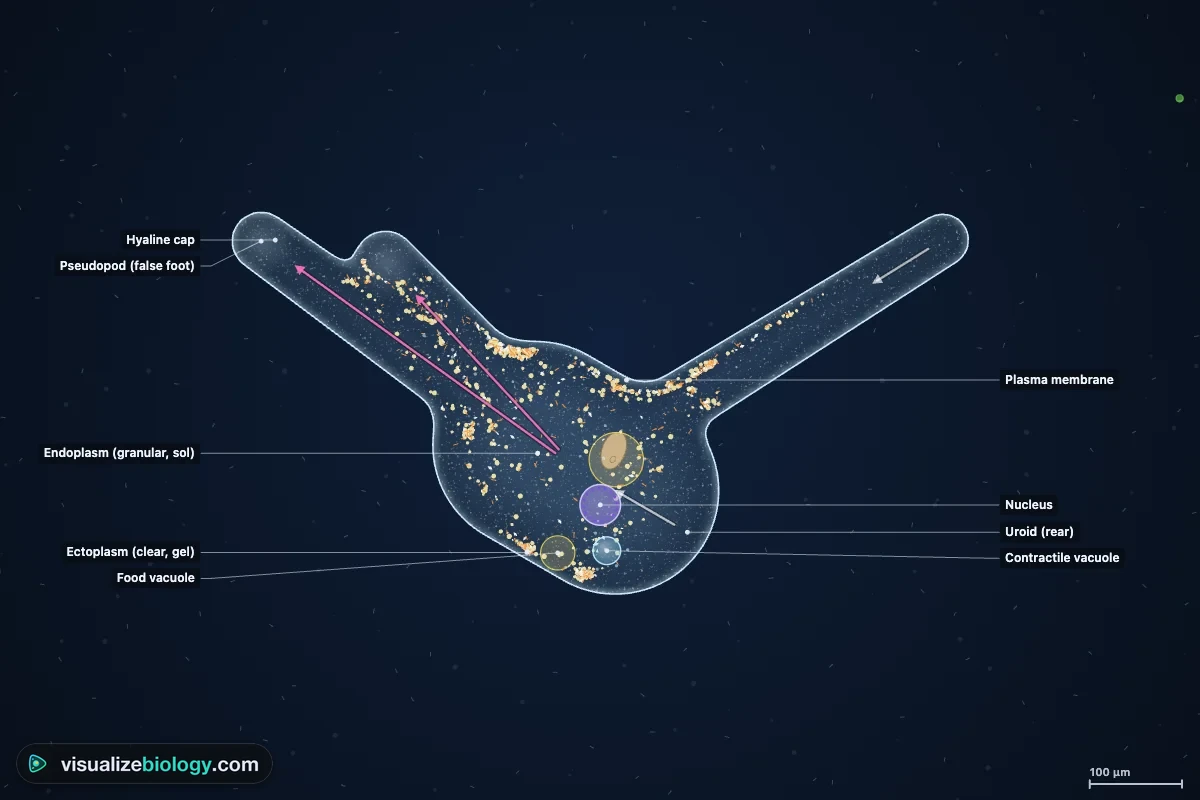
| Part | What it is | Function |
|---|---|---|
| Plasma membrane | A thin, flexible, partially permeable membrane | Controls what enters and leaves; oxygen, carbon dioxide and water pass across it |
| Pseudopodia (false feet) | Temporary, blunt bulges of cytoplasm | Movement (amoeboid movement) and catching food |
| Ectoplasm | The clear, firm (gel) outer layer of cytoplasm | Gives the cell its shape at that moment; forms the walls of a pseudopod |
| Endoplasm | The granular, fluid (sol) inner cytoplasm | Holds the organelles; streams forward into growing pseudopodia |
| Nucleus | A disc-shaped nucleus about 45 µm across | Holds the DNA and controls the cell; divides by mitosis in binary fission |
| Contractile vacuole | A clear, fluid-filled vacuole near the rear | Collects excess water and pumps it out (osmoregulation) |
| Food vacuoles | Membrane sacs around swallowed prey | Digest food with enzymes from lysosomes |
| Uroid | The wrinkled tail end | Contracts to push cytoplasm forward; waste leaves here |
| Hyaline cap | Clear cytoplasm at the tip of a growing pseudopod | Where new membrane and gel form as the pseudopod advances |
How does an amoeba move? Pseudopodia and the sol–gel cycle
An amoeba moves by amoeboid movement. It pushes out a pseudopod, and its cytoplasm flows in after it. The inner endoplasm is a runny sol; the outer ectoplasm is a stiff gel. At the back of the cell (the uroid) gel turns back into sol and the actin and myosin of the cortex contract, squeezing the sol forward, rather like toothpaste in a tube. At the front, in the fountain zone, the sol spreads out under the membrane and sets into gel, building the wall of the pseudopod as it grows. The front of Amoeba proteus advances only about 1 to 2 µm per second at room temperature, while the endoplasm streams two to three times faster. In the simulator the granules really do this: watch one stream forward, stop under the membrane at the tip, and stay put until the back of the cell reaches it. The same kind of movement is used by white blood cells as they crawl through tissues.
How does an amoeba eat? Phagocytosis and the food vacuole
Amoebae have holozoic nutrition: they take in solid food and digest it inside the cell. Chemicals leaking from a prey organism make the amoeba put out pseudopodia on that side (positive chemotaxis). The pseudopodia flow round the prey into a food cup, close behind it, and the prey ends up in a food vacuole inside the cell. This is phagocytosis, a kind of endocytosis. Lysosomes then fuse with the vacuole, pumping in acid and digestive enzymes: the vacuole's pH falls to about 4.5 within half an hour, the prey dies and is broken down, and the small soluble molecules are absorbed into the cytoplasm. The vacuole shrinks and returns to neutral, and hours later the undigested remains are pushed out at the uroid (egestion). Stain the prey with neutral red to watch the vacuole turn red as it becomes acid.
- Ingestion: pseudopodia surround the prey and enclose it in a food vacuole.
- Digestion: lysosomes add acid and enzymes to the food vacuole.
- Absorption: digested food diffuses into the cytoplasm.
- Assimilation: the cell uses it for energy and growth.
- Egestion: undigested waste is expelled at the back of the cell.
The contractile vacuole and osmoregulation
The cytoplasm of a freshwater amoeba is more concentrated than the pond water around it (about 0.10 osmol/L, a water potential of about −250 kPa, against almost zero outside). So water keeps entering the cell by osmosis. The contractile vacuole collects this water from small vesicles around it, swells, then fuses with the membrane and empties the water out, again and again. Pumping uses energy from ATP. In this model the vacuole of a 20 °C amoeba in distilled water empties about 3 times a minute, slowing as the outside solution gets stronger and stopping when it matches the cytoplasm. Put the amoeba in seawater and water leaves instead: the cell shrinks and its surface crinkles. Block the contractile vacuole and the amoeba swells and bursts in about an hour. This is an example of homeostasis in a single cell.
How does an amoeba breathe? Gas exchange and respiration
An amoeba has no lungs or gills. Oxygen dissolved in the water diffuses in across its whole membrane, down a concentration gradient, and carbon dioxide diffuses out. Inside, mitochondria use the oxygen in aerobic respiration: glucose + oxygen → carbon dioxide + water, releasing energy as ATP. Diffusion is fast enough only over short distances, so a single cell must have a large surface area to volume ratio. A sphere's SA:V is 3 ÷ radius: double the radius and the ratio halves. In this model the centre of a round cell more than about 0.6 mm across would run out of oxygen. Amoeba proteus avoids this by being flat as it crawls, about 40 µm thick, so no part of it is far from the surface. Its waste ammonia also leaves by diffusion. Try the agar cube lab to see why larger cubes are reached more slowly.
| Cube side | Surface area | Volume | SA : V | Reached by acid in 10 min (model) |
|---|---|---|---|---|
| 1 cm | 6 cm² | 1 cm³ | 6 : 1 | 87 % |
| 2 cm | 24 cm² | 8 cm³ | 3 : 1 | 57 % |
| 3 cm | 54 cm² | 27 cm³ | 2 : 1 | 41 % |
How does an amoeba reproduce? Binary fission
Amoebae reproduce asexually. When a well-fed Amoeba proteus has grown to about twice its size (about every two days at 20 °C) it divides by binary fission, which takes about an hour:
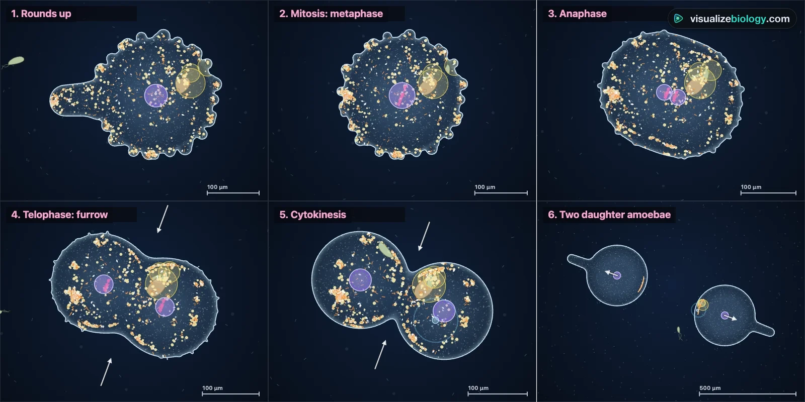
- The amoeba stops feeding, pulls in its pseudopodia and rounds up; its surface becomes covered in small knobs and the contractile vacuole disappears.
- The nucleus divides by mitosis (karyokinesis): its many small chromosomes line up and separate.
- The cell lengthens and a furrow pinches the cytoplasm in two (cytokinesis).
- Each daughter puts out pseudopodia, they crawl apart, and the last strand between them snaps.
The two daughter amoebae are genetically identical to each other and to the parent. Under good conditions the population doubles every generation, so a culture grows exponentially until food runs short. Some amoebae, such as Acanthamoeba and Entamoeba, survive drying or cold as cysts: the cell rounds up, secretes a tough wall, and its nucleus divides several times inside. When conditions improve the cyst bursts and releases many small amoebae. This is multiple fission, the textbook "encystment" of Amoeba. (Amoeba proteus itself is not known to form cysts.)
What does the nucleus do? The classic microsurgery experiment
If an amoeba is cut in two with a fine glass needle, both halves round up and close their membrane. The half with the nucleus soon crawls, feeds, grows and divides normally. The half without a nucleus can still move for a while, but it cannot digest food properly, grow or divide, and it dies within days. The experiment shows that the nucleus controls the cell: its DNA carries the instructions for making the proteins and enzymes the cell needs. Try it with the 🔪 Cut tool.
Amoeba under a microscope
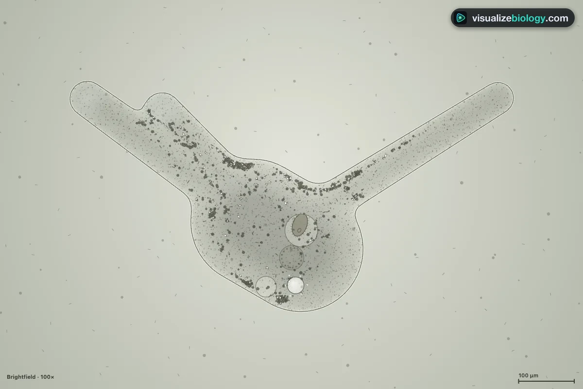
Amoebae settle on the bottom of a culture, so take your drop from there and support the coverslip so they are not crushed. At 40× an amoeba is a grey, granular, irregular blob that slowly changes shape; close down the condenser iris for more contrast. At 100× you can see pseudopodia, the clear ectoplasm, granular endoplasm streaming, the nucleus, the contractile vacuole filling and emptying, and food vacuoles. The Microscope look in this simulator and the virtual microscope's pond water slide show what to look for.
Amoeba vs paramecium vs euglena
| Amoeba | Paramecium | Euglena | |
|---|---|---|---|
| Shape | No fixed shape | Fixed slipper shape (pellicle) | Fixed spindle shape (pellicle) |
| Movement | Pseudopodia | Cilia | One flagellum |
| Nutrition | Heterotroph: phagocytosis anywhere on the surface | Heterotroph: food through the oral groove | Photosynthesis with chloroplasts; heterotroph in the dark |
| Water balance | One contractile vacuole | Two star-shaped contractile vacuoles | One contractile vacuole |
| Reproduction | Binary fission (multiple fission in cysts) | Binary fission; conjugation | Longitudinal binary fission |
Amoebas and human health
Most amoebae are harmless, but a few cause disease. Entamoeba histolytica causes amoebic dysentery when its cysts are swallowed in contaminated water or food. Naegleria fowleri, the so-called brain-eating amoeba, lives in warm fresh water and very rarely infects people when water goes up the nose. Acanthamoeba can infect the eye of contact lens wearers. Your own white blood cells (macrophages and neutrophils) crawl like amoebae and destroy bacteria by phagocytosis, just as an amoeba eats its prey. This simulator is an educational model, not medical advice.
Experiments and course links
| Lab | What you find out | Course links |
|---|---|---|
| Parts and size of an amoeba | Label the parts, measure the length, work out the magnification of a drawing | NGSS MS-LS1-1, MS-LS1-2; GCSE microscopy |
| Contractile vacuole and water potential | Count emptyings per minute in sucrose solutions; find the isotonic point | AP Biology 2.8; NGSS HS-LS1-3; A-level |
| Phagocytosis and food vacuole pH | Track a vacuole with neutral red as lysosomes make it acid | AP Biology 2.7; NCERT Class 10 Life Processes |
| Speed and temperature | Track the path, calculate speed and Q₁₀ | MS-LS1-1; enzymes and temperature |
| Agar cubes: why are cells small? | Surface area to volume ratio and diffusion | AP Biology 2.6; GCSE 4.1.3.1 |
| Population growth | Count cultures, plot a log graph, find the doubling time | Binary fission; ecology; NCERT Class 10 |
| Microsurgery | Which half of a cut amoeba survives, and why | NGSS MS-LS1-2; role of the nucleus |
Who uses this virtual amoeba?
Students use it to learn the parts of an amoeba and how a single cell carries out every life process, and to practice magnification, speed, osmosis and SA:V calculations with worked answers. Teachers project the 3D amoeba, pause it on each stage of binary fission, and assign the labs when there is no live culture or not enough microscopes. It is an educational model built from published measurements, it needs no login, and it runs on school Chromebooks.
Frequently asked questions
How does an amoeba move?
An amoeba moves by amoeboid movement: it pushes out temporary bulges of cytoplasm called pseudopodia (false feet). Fluid endoplasm (sol) streams forward into the growing pseudopod and turns into stiff ectoplasm (gel) at its tip, while at the back the gel turns back into sol and contracts, squeezing the cytoplasm forward. Actin and myosin drive the flow. Amoeba proteus crawls at about 1 to 2 micrometers per second.
How does an amoeba eat?
By phagocytosis. Pseudopodia flow around a bacterium, alga or small protist and close into a food cup, which pinches off inside the cell as a food vacuole. Lysosomes fuse with the vacuole and add acid and digestive enzymes, so its pH falls to about 4.5 and the food is broken down. The nutrients are absorbed into the cytoplasm and the undigested waste is pushed out at the back of the cell (egestion). This is holozoic nutrition.
What is the function of the contractile vacuole in an amoeba?
It removes excess water (osmoregulation). The cytoplasm of a freshwater amoeba is more concentrated than the pond water around it, so water keeps entering by osmosis. The contractile vacuole collects this water, swells and then empties it out through the cell membrane, using energy from ATP. If it stopped working, the amoeba would swell and burst. Marine amoebae, whose surroundings are as concentrated as their cytoplasm, have small or no contractile vacuoles.
How does an amoeba reproduce?
Asexually, by binary fission. When the amoeba has grown to about twice its size it stops, pulls in its pseudopodia and rounds up; the nucleus divides by mitosis, then the cytoplasm divides and two genetically identical daughter amoebae crawl apart. Amoeba proteus divides about every two days at 20 °C when well fed. Some amoebae survive harsh conditions as cysts and divide many times inside the cyst (multiple fission).
How does an amoeba breathe?
An amoeba has no lungs or gills: oxygen dissolved in the water diffuses in across its whole cell membrane, and carbon dioxide diffuses out. Inside, mitochondria use the oxygen for aerobic respiration, releasing energy as ATP. This works because a single small, flattened cell has a large surface area compared with its volume, so no part of it is far from the surface.
Is an amoeba a plant, an animal or a protist?
Neither a plant nor an animal: an amoeba is a single-celled eukaryote, traditionally placed in the kingdom Protista as a protozoan. It has a true nucleus and membrane-bound organelles, so it is not a prokaryote like bacteria. Modern classification puts Amoeba proteus in the group Amoebozoa. Because it is one cell, it carries out every life process (movement, nutrition, respiration, excretion, response, growth and reproduction) by itself.
Explore related simulators
Find amoebae, Paramecium and Euglena in pond water with the Virtual Microscope, see what water does to plant and animal cells in the Osmosis Lab, follow the nucleus of a dividing cell stage by stage in the Mitosis Simulator, and see the DNA that the nucleus holds in the DNA structure 3D model. The oxygen an amoeba uses comes from photosynthesis, shown in the Photosynthesis Lab. For something that is not alive in the same sense, compare the amoeba with the viruses of the Virus Virtual Lab: no cell, no metabolism, copied only inside a host. The bacteria an amoeba eats swim, divide and form spores in the Virtual Bacteria Simulator.
Sources: OpenStax Biology 2e, chapter 23 Protists; Jeon (ed.), The Biology of Amoeba (Academic Press, 1973); Rogerson, Canadian Journal of Zoology 58 (1980), on the generation time of Amoeba proteus (44–84 h at 20 °C); Allen and Allen, Annual Review of Biophysics and Bioengineering 7 (1978), on cytoplasmic streaming in amoeboid movement; College Board, AP Biology Course and Exam Description, Unit 2. Speeds, rates and sizes are an educational model based on these sources.
Updated