
Virus Virtual Lab
Build a virus, cut it open and name its parts, then infect a cell and watch it take over: the lytic and lysogenic cycles, HIV, flu and 14 more viruses. Block each step with a drug, then run five virus labs.
Live math
Lab notebook
Every infection you run (what was blocked, how far it got, how many virions it made) and every measurement in the labs is written here. Export a CSV, or turn the session into a PDF lab report.
| # | Lab / virus | Conditions | Measurement | Result |
|---|
Concept library
Click "New problem" to begin.
Click "Start quiz" for 5 questions: the parts of a virus, replication, the lytic and lysogenic cycles, classification, lab calculations and antivirals.
1 Overview
Simulate has 17 viruses (choose one with Virus in the top bar). The left card shows one Virion you can turn, cut open and label, or the Infection: the virus attacking its host cell (a bacterium, an animal cell or a plant cell) step by step. The right card shows the Genome (the Baltimore classification), the Cycle (the replication cycle, with the live moment marked), Graph and Size. Experiment has five labs that end in a PDF lab report, and Quiz has a practice quiz with worked solutions and a 5-question quiz.
2 Getting started
- The virus opens in 3D on a computer or tablet and in 2D on a phone. The 2D | 3D switch changes it and remembers your choice.
- In Virion, drag to turn the virus, switch on Cutaway to see inside, and hover or click any part to read what it does. T4 can contract its tail (Record panel).
- Press Infect (or switch to Infection and press ▶). Teach gives every step a few seconds; the other speeds run in the virus's own time (a T4 cycle takes about 25 minutes, HIV about a day).
- The caption says what is happening; Next step (N) jumps ahead, and the strip and the Cycle wheel let you click any step.
- Open Status (S) for live readings: the step, the time since infection, genome copies and new virions.
3 The infection panel
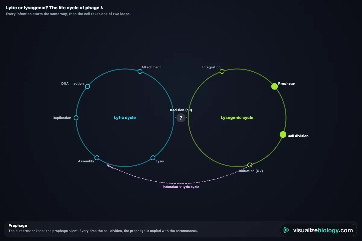
- Phage λ can take the lytic or the lysogenic path. Let the cell decide (more phages per cell and starved cells favor lysogeny), or choose a path. A lysogen keeps the prophage until you shine UV light.
- Herpes simplex can infect a skin cell (lytic) or hide in a neuron (latency).
- Temperature changes how fast a phage cycle runs (Q₁₀ ≈ 2); below about 16 °C or above about 44 °C its host stops growing.
- Infect a cell starts again with a fresh host.
4 Block it on purpose
Antibodies from a vaccine, a cell without the receptor, or an antiviral drug stop the virus at one step: maraviroc (entry), AZT and tenofovir (reverse transcription), dolutegravir (integration), protease inhibitors (maturation), oseltamivir (release), baloxavir, remdesivir and Paxlovid (synthesis), acyclovir (DNA copying). The cycle runs up to that step and stops, and the coach explains why. Try an antibiotic too: it does nothing, because viruses have none of the bacterial structures antibiotics attack.
5 Genome, Cycle, Graph and Size
- Genome: the seven Baltimore groups and the route each genome takes to mRNA; this virus's group is lit. Click a group to see its examples.
- Cycle: the six steps (attachment, entry, uncoating, replication and synthesis, assembly, release) plus reverse transcription, integration, maturation or latency where they apply; for λ, the lytic and lysogenic loops. Click a step to jump there.
- Graph: the one-step growth curve, MOI and the Poisson distribution, λ's decision, plaque counts, mutations per genome, herd immunity and latent period against temperature. Your lab results appear as gold dots.
- Size: this virus on a log scale from an atom to a cheek cell, and drawn to scale beside E. coli.
6 Experiment mode and the lab report
Five labs: the plaque assay (make serial dilutions, plate them, incubate, count the plaques and calculate the titer), the one-step growth curve (sample an infected culture over time and find the latent period and burst size), lytic or lysogenic (infect at different MOIs and score lysogens), MOI and the Poisson distribution (count surviving cells), and identifying four mystery viruses from an electron micrograph and four tests. Each lab shows its aim, what you need and a real-lab safety note; the method ticks off as you work, a conclusion question checks your own result, and Create lab report builds a PDF.
7 Quiz
The practice quiz asks you to name parts on a picture of a virus, order the steps of replication, tell lytic from lysogenic, classify genomes, and calculate titers, dilutions, MOI, burst size, sizes from electron micrographs, mutations per genome and herd immunity thresholds, each with a worked solution. Take the quiz gives five mixed questions and a star rating.
8 Keyboard and gestures
- Space plays or pauses, N goes to the next step, V and I switch between Virion and Infection, C toggles the cutaway, S opens Status, G shows the graph.
- Scrolling over a view always scrolls the page. To zoom, hold Ctrl (⌘ on a Mac) and scroll, or pinch on a trackpad. On a phone one finger scrolls the page; turn the 3D virus with two fingers.
- Right-click a view to save it as an image or copy your results.
Virus virtual lab: the structure of viruses and how they replicate
A virus is a package of genes in a protein coat that can only be copied inside a living cell. It has no metabolism and no ribosomes of its own, so it hijacks the host's. This free virus virtual lab lets you build seventeen viruses in 3D, cut them open and name their parts, then follow each one as it infects its host cell: a bacterium, a human cell or a plant cell. You can stop any step with a drug, watch phage lambda choose between the lytic and lysogenic cycles, and run the classic phage labs used in high school and college courses: the plaque assay, the one-step growth curve and the multiplicity of infection.
- Viruses
- T4 and λ phages, TMV, influenza A, HIV-1, SARS-CoV-2, poliovirus, rhinovirus, adenovirus, herpes simplex 1, HPV, measles, rabies, Ebola, rotavirus, parvovirus B19, hepatitis B
- Hosts
- E. coli bacteria, human and animal cells (including a neuron), tobacco leaf cells
- Processes
- Attachment, entry, uncoating, replication and synthesis, assembly, release by lysis or budding; reverse transcription, integration, lysogeny and induction, latency
- Views
- 3D or 2D virion with cutaway and labels; the infection; the Baltimore classification; the replication cycle; graphs; a size scale
- Labs
- Plaque assay, one-step growth curve, lytic or lysogenic, MOI and the Poisson distribution, identify a mystery virus
- Modes
- Simulate, Experiment (5 labs) and Quiz
- Runs on
- Any modern browser, including Chromebooks, iPads and phones. 3D on larger screens, 2D on phones.
- Price
- Free, with no account or download
Is a virus living or nonliving?
Viruses sit on the border of life. Like living things, they carry genes made of DNA or RNA, they reproduce and they evolve by natural selection. But a virus is not a cell: it has no cytoplasm, ribosomes or enzymes for making energy, it does not grow, and outside a host it is an inert particle (a virion). It can only be copied inside a living cell, so viruses are obligate intracellular parasites. That is why they are not placed in any of the three domains of life.
| Trait of life | Cells | Viruses |
|---|---|---|
| Made of cells | Yes | No: a genome in a protein coat |
| DNA and RNA | Both | One or the other |
| Metabolism and ribosomes | Yes | No: they use the host's |
| Reproduce | On their own | Only inside a host cell |
| Grow, respond, keep homeostasis | Yes | No |
| Evolve | Yes | Yes, often fast |
Parts of a virus
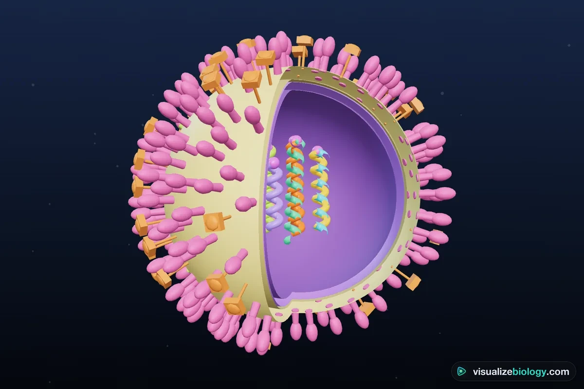
Switch the lab to Virion and turn on the labels for a labeled diagram of any virus; turn on Cutaway to see inside.
| Part | What it is | Function |
|---|---|---|
| Genome | DNA or RNA; single- or double-stranded; one piece or several segments | Carries the genes for the virus's proteins |
| Capsid | A protein shell built from many identical capsomeres | Protects the genome and, in naked viruses, attaches to the host |
| Envelope | A lipid bilayer taken from the host cell as the virus buds out | Lets the virus fuse with the next cell; makes it sensitive to soap and alcohol |
| Spikes (glycoproteins) | Viral proteins in the envelope, such as hemagglutinin, gp120 and the coronavirus spike | Bind receptors on the host cell; the main target of vaccines |
| Matrix | A protein layer under the envelope | Links the envelope to the nucleocapsid and shapes the virion |
| Enzymes | Reverse transcriptase, integrase, RNA polymerase… | Jobs the host cannot do, such as copying RNA |
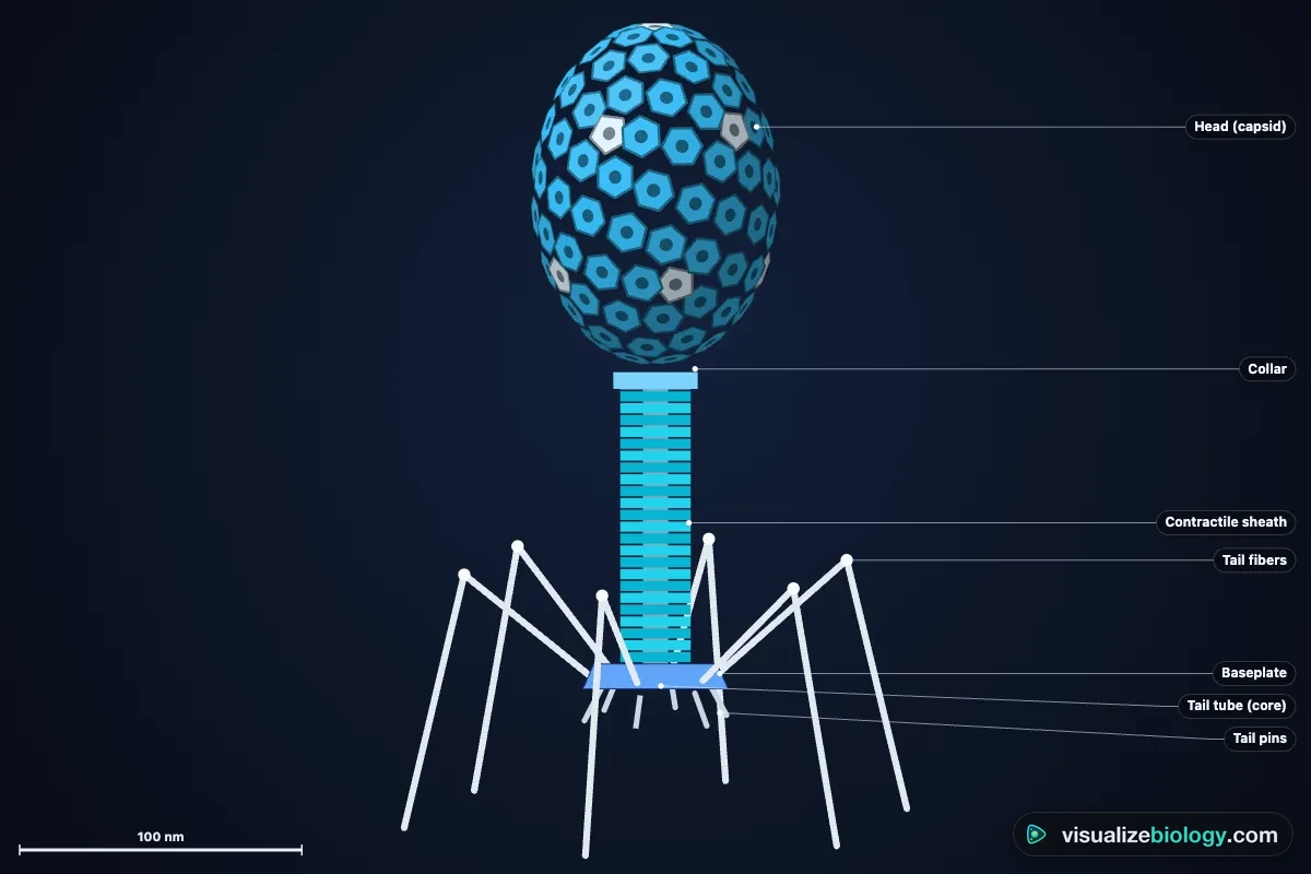
| Tail, sheath, baseplate, tail fibers | The injection machine of tailed phages | Find the bacterium and inject the DNA |
Virus shapes: helical, icosahedral, complex and enveloped
Helical capsids are rods of identical proteins wound round the genome: tobacco mosaic virus has 2130 of them, 16.3 per turn. Icosahedral capsids have 20 triangular faces; they are built on a lattice with exactly 12 five-fold corners, so a capsid of triangulation number T has 10T + 2 capsomeres (32 for poliovirus, 72 for HPV, 252 for adenovirus). Complex viruses such as T4 combine an icosahedral head with a helical tail. Any of these can be enveloped (influenza, HIV, SARS-CoV-2, herpes) or non-enveloped, also called naked (polio, adenovirus, HPV). Enveloped viruses are fragile outside the body; naked viruses survive on surfaces and in the gut.
How do viruses replicate? The six steps
- Attachment: proteins on the virus bind specific receptors on the host cell (HIV's gp120 binds CD4 and CCR5; the SARS-CoV-2 spike binds ACE2). Receptors decide the host range.
- Entry (penetration): phages inject their DNA; enveloped viruses fuse with the membrane; others are swallowed by endocytosis.
- Uncoating: the capsid comes apart and releases the genome.
- Replication and synthesis: the genome is copied and viral mRNA is translated on the host's ribosomes.
- Assembly: capsid proteins self-assemble round new genomes.
- Release: by lysis (the cell bursts) or budding (the virus wraps itself in the cell membrane).
Lytic vs lysogenic cycle
| Lytic cycle | Lysogenic cycle | |
|---|---|---|
| Viral DNA | Stays separate and is copied many times | Inserted into the host chromosome as a prophage |
| Host cell | Taken over and killed | Lives and divides normally; every daughter cell carries the prophage |
| New viruses | About 100–150 per cell within about 25–50 minutes | None while the prophage is silent |
| Kept quiet by | — | The cI repressor protein |
| Ends with | Lysis | Induction (UV light or DNA damage) then the lytic cycle |
| Examples | T4 (virulent phage); λ when it chooses lysis | λ (temperate phage); similar latency in herpes and HIV |
What decides? In phage λ the cII protein: when several phages infect one cell, or the cell is starved, cII builds up and switches on the repressor, so lysogeny wins. It is a sensible bet: many phages per cell means few uninfected cells are left to infect. In the lab, set the number of phages per cell and watch the decision change.
The Baltimore classification
Every virus must make mRNA for the host's ribosomes. David Baltimore grouped viruses by the route their genome takes to mRNA.
| Group | Genome | Route to mRNA | Examples |
|---|---|---|---|
| I | dsDNA | Transcribed like the cell's own genes | T4, λ, adenovirus, herpes, HPV |
| II | ssDNA | Made double-stranded first | Parvovirus B19 |
| III | dsRNA | Viral RNA polymerase inside the capsid | Rotavirus |
| IV | (+)ssRNA | The genome is mRNA | Poliovirus, rhinovirus, SARS-CoV-2, TMV |
| V | (−)ssRNA | Copied by a polymerase carried in the virion | Influenza, measles, rabies, Ebola |
| VI | ssRNA with reverse transcription | RNA → DNA → provirus → mRNA | HIV |
| VII | dsDNA with reverse transcription | DNA → RNA → DNA | Hepatitis B |
Retroviruses and the HIV life cycle
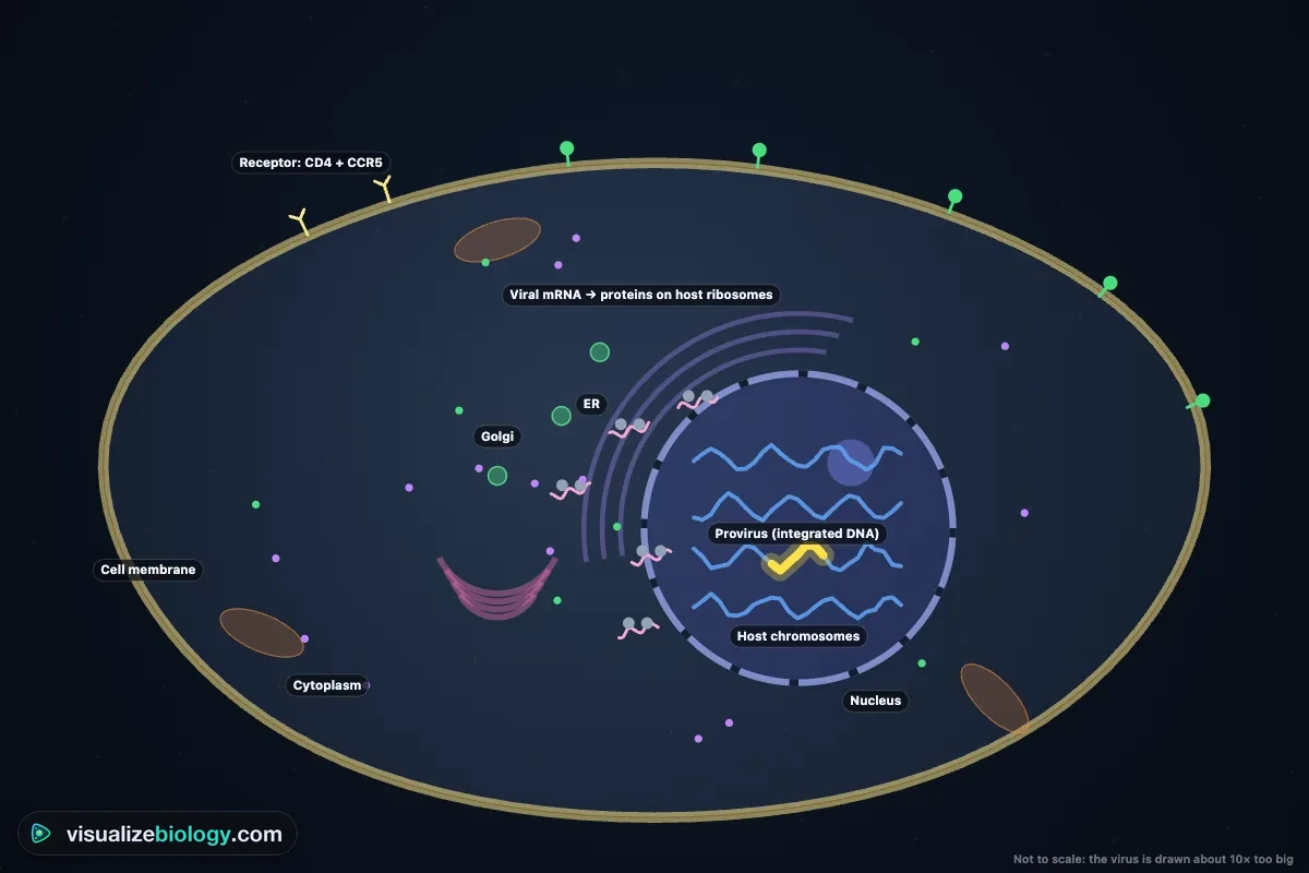
HIV is a retrovirus. Its gp120 spikes bind CD4 and a co-receptor on helper T cells; gp41 fuses the membranes. Inside the cone-shaped capsid, reverse transcriptase copies the RNA genome into DNA, and integrase splices that DNA into a chromosome, where it stays for the life of the cell as a provirus. The cell then makes viral RNA and proteins, new virions bud from the membrane, and the viral protease cuts the Gag protein so they mature. Each step is a drug target in antiretroviral therapy: entry inhibitors, reverse transcriptase inhibitors such as AZT and tenofovir, integrase inhibitors and protease inhibitors. Reverse transcriptase has no proofreading, making about 2 × 10⁻⁵ errors per base, roughly one new mutation in every genome it copies, which is why HIV evolves drug resistance and why combinations of drugs are used.
Antigenic drift vs antigenic shift
Influenza A changes in two ways. Antigenic drift is the slow build-up of point mutations in the genes for hemagglutinin (HA) and neuraminidase (NA), so antibodies from last year's infection or vaccine fit less well; it is why the flu vaccine is updated every year. Antigenic shift is sudden: when two strains infect the same cell, their eight RNA segments can be swapped (reassortment, up to 2⁸ = 256 combinations), producing a new subtype such as H1N1 or H3N2 that few people are immune to. Shifts caused the pandemics of 1957, 1968 and 2009.
Virus vs bacteria
| Virus | Bacterium | |
|---|---|---|
| Size | 20–300 nm (Ebola filaments about 1 µm) | 1–5 µm |
| Structure | Genome in a capsid, sometimes an envelope | A prokaryotic cell: wall, membrane, cytoplasm, ribosomes |
| Genetic material | DNA or RNA | DNA (and RNA) |
| Reproduction | Only inside a host cell | Binary fission, on its own |
| Seen with | An electron microscope | A light microscope |
| Treated with | Antivirals and vaccines | Antibiotics (and vaccines for some) |
The plaque assay: counting viruses
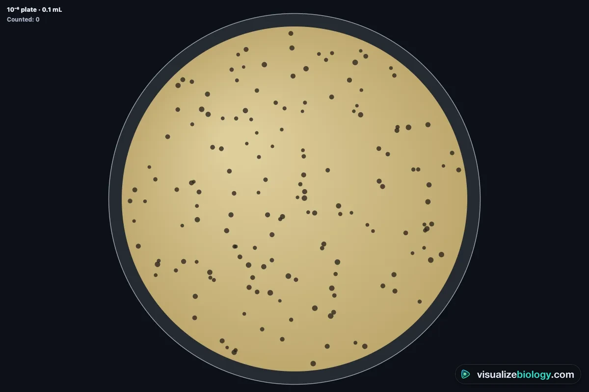
Phages are counted by the holes they make in a lawn of bacteria. A stock is diluted tenfold again and again, a small volume of each dilution is mixed with bacteria in soft agar and poured onto a plate, and overnight each infectious phage starts a round of infection that spreads into a clear plaque. Count a plate with 30 to 300 plaques, then:
titer (PFU/mL) = plaques ÷ (dilution × volume plated)
For example, 122 plaques from 0.1 mL of the 10⁻⁶ dilution: 122 ÷ (10⁻⁶ × 0.1 mL) = 1.22 × 10⁹ PFU/mL. In a one-step growth curve the same counts, taken every few minutes after a single round of infection, show the eclipse and latent periods and the burst size: about 25 minutes and 150 phages per cell for T4 at 37 °C. The multiplicity of infection (MOI) is the ratio of phages to cells; because phages land on cells at random, the fraction of cells left uninfected is e−MOI, so an MOI of 3 is needed to infect 95 % of them.
Vaccines, antivirals, prions and viroids
Vaccines train the immune system to make antibodies and memory cells against a virus's surface proteins before infection, so the virus is neutralized at attachment. When enough people are immune, a virus cannot spread: the herd immunity threshold is 1 − 1/R₀, about 95 % for measles. Antivirals block steps that the virus does differently from the cell, such as reverse transcription. Antibiotics do not work on viruses. Two other acellular agents are smaller still: viroids, naked circles of RNA a few hundred bases long that infect plants, and prions, misfolded proteins with no genome at all that cause mad cow disease and Creutzfeldt–Jakob disease. This lab is an educational model, not medical advice.
Experiments and course links
| Lab | What you find out | Course links |
|---|---|---|
| Plaque assay | Make serial dilutions, count plaques, calculate the titer | OpenStax Microbiology 6.3; college microbiology lab |
| One-step growth curve | Latent period, eclipse and burst size of T4 | OpenStax Microbiology 6.2; AP Biology 6.4 |
| Lytic or lysogenic? | How MOI and starvation tip phage λ toward lysogeny | OpenStax Biology 21.2; AP Biology 6.7 |
| MOI and the Poisson distribution | Surviving fraction against MOI; e−MOI | College microbiology and virology |
| Identify the mystery virus | Shape, envelope, DNA or RNA, strandedness → Baltimore group | OpenStax Biology 21.1; NGSS HS-LS1-1 |
Who uses this virus lab?
Students use it to learn the parts of a virus, the steps of viral replication and the lytic and lysogenic cycles, and to practice titer, MOI and burst-size calculations with worked answers. Teachers project the 3D viruses, pause an infection at each step, show how each HIV drug works, and assign the phage labs when there is no microbiology lab or not enough time. It is an educational model built from published measurements, it needs no login, and it runs on school Chromebooks.
Frequently asked questions
What is the difference between the lytic and lysogenic cycle?
In the lytic cycle a virus takes over the host cell at once: it copies its genome, makes new virions and bursts (lyses) the cell, killing it and releasing about 100 new viruses within an hour for a phage. In the lysogenic cycle the viral DNA is inserted into the host chromosome as a prophage and is copied every time the cell divides, without harming it. Stress such as UV light can later induce the prophage to leave the chromosome and enter the lytic cycle. Phage lambda can do either; phage T4 is lytic only.
What are the parts of a virus?
Every virus has a genome (DNA or RNA, never both) inside a protein coat called the capsid, which is built from repeating protein units called capsomeres. The genome and capsid together are the nucleocapsid. Many animal viruses also have an envelope, a lipid membrane taken from the host cell, studded with glycoprotein spikes that bind receptors on the next cell. Some viruses carry enzymes such as reverse transcriptase or RNA polymerase. Bacteriophages like T4 add a tail: a collar, a contractile sheath round a tail tube, a baseplate and tail fibers.
Is a virus living or nonliving?
Most biologists place viruses on the border of life. A virus has genes and evolves by natural selection, but it is not made of cells, has no metabolism or ribosomes, does not grow, and cannot reproduce on its own: it can only be copied inside a living host cell, using the cell's ribosomes, enzymes and energy. Outside a cell a virion is an inert particle. For that reason viruses are called obligate intracellular parasites and are not placed in any of the three domains of life.
How do viruses replicate?
In six steps. Attachment: proteins on the virus bind specific receptors on the host cell. Entry: the genome is injected (phages) or the virus fuses with the membrane or is taken in by endocytosis. Uncoating: the capsid releases the genome. Replication and synthesis: the cell copies the viral genome and makes viral proteins on its own ribosomes. Assembly: new capsids form round copies of the genome. Release: the new virions leave by bursting the cell (lysis) or by budding through its membrane, which gives them an envelope.
How is a virus different from a bacterium?
A bacterium is a living prokaryotic cell, about 1 to 5 micrometers long, with a cell wall, a membrane, ribosomes and both DNA and RNA; it grows and divides on its own and can be killed by antibiotics. A virus is about 20 to 300 nanometers across, roughly 10 to 100 times smaller, has only DNA or RNA in a protein coat, has no ribosomes or metabolism, and can only be copied inside a host cell. Antibiotics do not work against viruses; antiviral drugs and vaccines do.
How do you calculate the titer from a plaque assay?
Count the plaques on a plate that has between 30 and 300 of them, then divide by the dilution of that plate and by the volume plated in milliliters: titer (PFU/mL) = number of plaques ÷ (dilution × volume plated). For example, 122 plaques from 0.1 mL of a 10⁻⁶ dilution give 122 ÷ (10⁻⁶ × 0.1) = 1.22 × 10⁹ plaque-forming units per mL.
Explore related simulators
Translate a viral gene yourself, and see why one base can turn a missense into a frameshift, in the Interactive Codon Chart and DNA to Protein Translator. See the DNA that most viruses carry in the DNA structure 3D model, watch the host cell's own copying machinery at work in the Mitosis Simulator, find bacteria and protists under the Virtual Microscope, and meet a single-celled organism that is alive in every sense in the Virtual Amoeba Simulator. Viral recombination echoes crossing over in the Meiosis Simulator. The bacteria that phages infect, and how they take up genes by transduction, are alive in the Virtual Bacteria Simulator.
Sources: OpenStax Biology 2e, chapter 21 Viruses; OpenStax Microbiology, chapter 6 Acellular Pathogens; Flint et al., Principles of Virology (4th ed., ASM Press); ICTV virus taxonomy; Ellis and Delbrück, Journal of General Physiology 22 (1939), on the one-step growth curve; Kourilsky, Molecular and General Genetics 122 (1973), and Zeng et al., Cell 141 (2010), on lysogeny and MOI; Sanjuán et al., Journal of Virology 84 (2010), on viral mutation rates; Caspar and Klug (1962) on capsid architecture; College Board, AP Biology Course and Exam Description, Units 6 and 7. Sizes, times and rates are an educational model based on these sources.
Updated