
Virtual Bacteria Simulator
Sixteen real bacteria, alive and to scale in 3D: zoom into a cell's wall, DNA and flagella, watch it swim, divide and fill a drop of broth, starve it into spores, feed it antibiotics or new genes. Then Gram stain, count and test bacteria in seven virtual labs.
Live math
Lab notebook
Every measurement you record, and every event in the culture (a conjugation, a burst, the first spore), is written here with its conditions. Export a CSV, or turn the session into a PDF lab report.
| # | Lab / sample | Conditions | Measurement | Result |
|---|
Concept library
Click "New problem" to begin.
Click "Start quiz" for 5 questions: the bacterial cell, types and the Gram stain, growth, energy, genetics and antibiotics.
1 Overview
Simulate shows living bacteria in a drop of broth or on an agar surface. Pick one of sixteen real species with Bacterium, and a topic with Show: the parts of a bacterium, movement, binary fission, the growth curve, colonies, respiration and fermentation, gene transfer, antibiotics, endospores or the Gram stain. Change the temperature, pH, oxygen and food, add antibiotics, donors or phages, starve them, boil or autoclave them. Beside them, Inside shows what no light microscope can (the cell envelope, the flagellar motor, DNA being copied, the electron transport chain, gene transfer, spore formation), Stain shows how the species looks after the Gram, acid-fast, endospore and capsule stains, Graph plots growth and the effect of temperature, pH and oxygen, and Types lists all sixteen bacteria. Experiment has seven labs that end in a PDF lab report, and Quiz has a practice quiz with worked solutions and a 5-question quiz.
2 Getting started
- The bacteria open in 3D on a computer or tablet and in 2D on a phone. The 2D | 3D switch changes it and remembers your choice. Textbook | Phase contrast changes the look: colored diagram, or living cells as they look at 1000× (no flagella: they are too thin to see).
- Cell | Field (or Z) zooms between one cell and the whole field of view. Hold Ctrl (⌘ on a Mac) and scroll to zoom further.
- Press ▶ (or Space). Growth runs at the speed you pick, from real time to one hour per second. Swimming is shown slowed so you can follow each cell: 4× slower in the Field view and about 40× slower in the Cell view (the view says how much, and gives the real speed, about 25 µm/s for E. coli).
- Click a cell to follow it, or a part to name it. The caption says what is happening now; the cell-cycle strip shows where the followed cell is in its life.
- Open Status (S) for live readings: what the cell is doing, the number of cells, the growth phase, the doubling time, how it makes energy and how much food is left.
3 Tools and panels
- 👆 Look names parts and follows cells; 📏 Measure measures in µm; 🍬 Food drops food that motile cells swim toward (chemotaxis) and that makes spores germinate; 🦠 Phage adds bacteriophages.
- Conditions: a drop of broth or an agar surface; rich, minimal or no food; temperature, pH and oxygen; light for the cyanobacterium; nitrate for anaerobic respiration.
- Antibiotics: nine drugs and a dose in multiples of the species' MIC. Wall drugs burst growing cells, ribosome drugs stop growth, ciprofloxacin makes filaments, polymyxin dissolves Gram-negative membranes. "Make some cells resistant" gives a few cells an R plasmid (red ring).
- Genes & survival: F⁺ and R-plasmid donors for conjugation, heat-killed smooth pneumococci for Griffith's experiment, phages, starve, feed, boil and autoclave.
4 Inside, Stain, Graph and Types
- Envelope: the Gram-positive, Gram-negative or acid-fast wall to scale, with the Gram stain played out on it. Flagellum: the rotary motor, run and tumble, and the five arrangements. Divide: binary fission of the cell you are following. Energy: glycolysis, the Krebs cycle and the electron transport chain in the membrane, or fermentation, with ATP per glucose. Genes: conjugation, transformation and transduction step by step. Spore: the seven stages of sporulation.
- Stain: a smear of the species at 1000× after the Gram, acid-fast, endospore or capsule stain.
- Graph: growth in the drop on a log scale with its phases; growth rate against temperature and pH; thioglycolate tubes; energy yield; sizes from a ribosome to a giant bacterium.
- Types: click any of the sixteen bacteria to study it.
5 Experiment mode and the lab report
Seven labs: Gram staining three unknowns, the growth curve of E. coli (OD₆₀₀ and generation time), serial dilution and plate count (CFU/mL), Kirby–Bauer antibiotic sensitivity, oxygen requirements in thioglycolate tubes with a catalase test, pGLO transformation (AP Biology Investigation 8) and the evolution of antibiotic resistance. Each shows its aim, what you need and a real-lab safety note. The method ticks off as you work, the bench shows the apparatus, the observe card shows what you look at (a smear, a plate, a tube), a conclusion question checks your result, and Create lab report builds a PDF with your results, figures, calculations and sources of error.
6 Quiz
The practice quiz covers the parts of a bacterial cell (with a picture), magnification, shapes and arrangements, the Gram stain, oxygen classes, generation time and CFU calculations, the growth curve, respiration and fermentation, gene transfer, pGLO efficiency and antibiotics, each with a worked solution. Take the quiz gives five mixed questions and a star rating.
7 Keyboard and gestures
- Space plays or pauses, N goes to the next stage of the cell cycle, Z switches Cell and Field, S opens Status, I and G switch to Inside and Graph, M picks the measuring tool and Esc goes back to Look.
- Scrolling over the bacteria always scrolls the page. To zoom, hold Ctrl (⌘ on a Mac) and scroll, or pinch. On a phone, one finger scrolls the page; turn the 3D view or zoom the 2D view with two fingers.
- Right-click a view to save it as an image or copy your results.
Bacteria: structure, types, growth and how they live
Bacteria are single-celled prokaryotes: living cells without a nucleus, usually 0.5 to 5 µm long. They were the first life on Earth and are still the most numerous: a gram of soil holds billions, and your own body carries about as many bacterial cells as human ones. This free virtual bacteria simulator shows sixteen real species alive and to scale. You can zoom into one cell's wall and DNA, watch it swim, divide and fill a drop of broth, starve it into spores, give it antibiotics or new genes, and then stain, count and test bacteria in seven virtual labs.
- Organisms
- Sixteen real bacteria, from E. coli and Staphylococcus to Borrelia, Mycobacterium tuberculosis and the cyanobacterium Anabaena
- Processes
- Swimming and chemotaxis, binary fission, the growth curve, respiration and fermentation, gene transfer, sporulation, antibiotic action and resistance
- Views
- 3D or 2D; textbook diagram or phase contrast; close-ups; stained smears; live graphs
- Labs
- Gram stain, growth curve, plate count, Kirby–Bauer, thioglycolate, pGLO transformation, evolution of resistance
- Modes
- Simulate, Experiment (7 labs) and Quiz
- Runs on
- Any modern browser, including Chromebooks, iPads and phones. 3D on larger screens, 2D on phones.
- Price
- Free, with no account or download
Parts of a bacterial cell
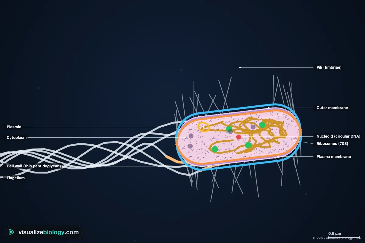
A bacterium is a prokaryotic cell: it has no nucleus and no membrane-bound organelles such as mitochondria or chloroplasts. Everything happens in the cytoplasm or on the plasma membrane.
| Part | What it is | Function |
|---|---|---|
| Capsule | A slimy layer of polysaccharide (not in every species) | Sticks to surfaces; hides the cell from white blood cells |
| Outer membrane | A second membrane with lipopolysaccharide (Gram-negative bacteria only) | A barrier to many antibiotics; LPS is endotoxin |
| Cell wall | Peptidoglycan: sugar chains cross-linked by peptides | Keeps the shape and stops the cell bursting; the target of penicillin |
| Plasma membrane | A phospholipid bilayer | Controls what enters and leaves; respiration and ATP synthesis happen here |
| Cytoplasm | A gel of water, enzymes and food molecules | Where metabolism and protein synthesis happen |
| Nucleoid | One circular chromosome, folded (E. coli: 4.6 million base pairs) | Carries the genes; there is no nuclear membrane |
| Plasmids | Small extra circles of DNA | Extra genes, such as antibiotic resistance; can pass between cells |
| Ribosomes (70S) | Smaller than ours (80S) | Make proteins; the target of tetracycline and erythromycin |
| Pili (fimbriae) | Short protein hairs | Attach to surfaces; a sex pilus passes DNA in conjugation |
| Flagella | Long helical filaments turned by a rotary motor | Swimming; driven by protons flowing into the cell |
Types of bacteria: shapes and arrangements
Bacteria are first described by their shape: cocci (spheres), bacilli (rods), vibrios (commas), spirilla (rigid spirals) and spirochetes (flexible spirals). The arrangement comes from how the cells divide and stay together: diplococci in pairs, streptococci in chains (one plane of division), staphylococci in grape-like clusters (random planes), tetrads in fours (two planes at right angles). Put a single cell on the agar surface in the simulator and watch its arrangement appear as it divides.
| Bacterium | Shape and arrangement | Gram | Oxygen | Known for |
|---|---|---|---|---|
| Escherichia coli | Rod, single | − | Facultative anaerobe | Gut; the lab workhorse; divides every 20 min |
| Staphylococcus aureus | Cocci in clusters | + | Facultative anaerobe | Skin infections, MRSA |
| Streptococcus pyogenes | Cocci in chains | + | Aerotolerant anaerobe | Strep throat |
| Bacillus subtilis | Rods, chains | + | Aerobe | Soil; endospores |
| Clostridium tetani | Rod with a terminal spore | + | Obligate anaerobe | Tetanus |
| Vibrio cholerae | Comma (vibrio) | − | Facultative anaerobe | Cholera; one polar flagellum |
| Borrelia burgdorferi | Spirochete | − | Microaerophile | Lyme disease, carried by ticks |
| Mycobacterium tuberculosis | Rod, cords | Acid-fast | Obligate aerobe | Tuberculosis; doubles every 15–20 h |
Gram-positive vs Gram-negative bacteria and the Gram stain
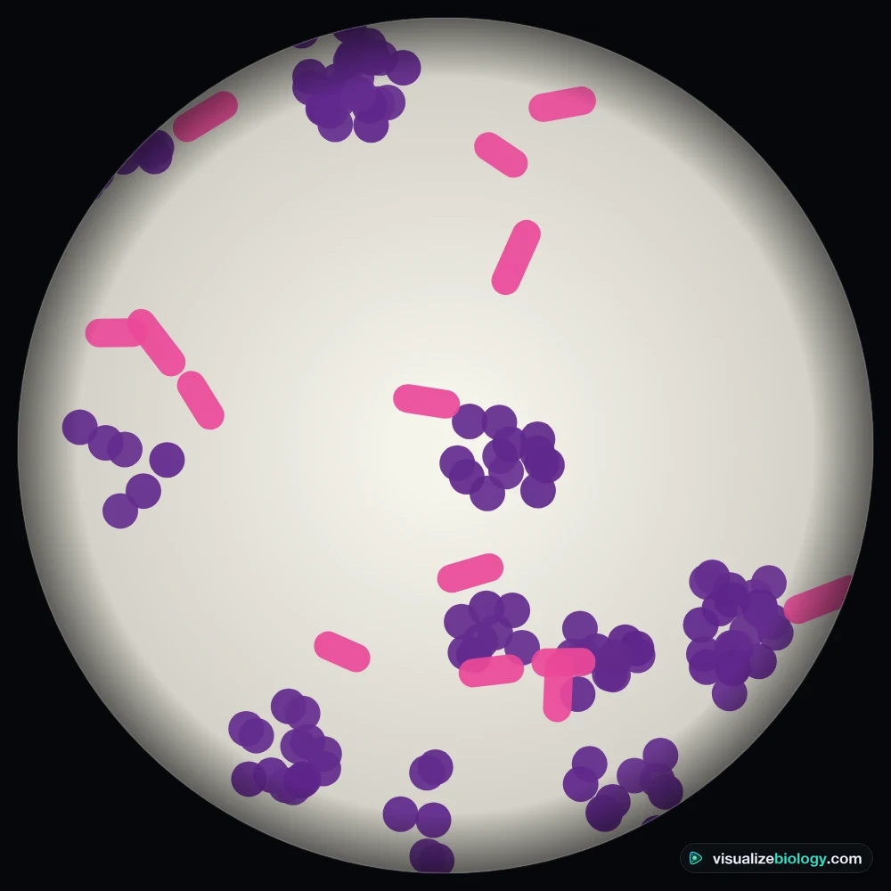
The Gram stain, invented by Hans Christian Gram in 1884, splits most bacteria into two groups by their cell envelope. Every cell first takes up purple crystal violet; iodine turns it into a large crystal violet–iodine complex. Alcohol then dehydrates the thick peptidoglycan of a Gram-positive wall and traps the complex inside, but it dissolves the outer membrane of a Gram-negative cell and washes the dye out of its thin wall. A pink counterstain, safranin, colors the decolorized cells. In the Gram stain lab you can see what goes wrong when a step is skipped: without iodine, or with too long in alcohol, Gram-positive cells look pink.
| Gram-positive | Gram-negative | |
|---|---|---|
| Color after the Gram stain | Purple | Pink |
| Peptidoglycan | Thick, 20–80 nm, with teichoic acids | Thin, 2–7 nm |
| Outer membrane | None | Yes, with lipopolysaccharide (endotoxin) and porins |
| Periplasm | Very small | A space between the two membranes |
| Penicillin and vancomycin | Usually work well | Often kept out by the outer membrane |
| Examples | Staphylococcus, Streptococcus, Bacillus, Clostridium, Listeria | E. coli, Salmonella, Vibrio, Pseudomonas, Helicobacter |
Mycobacteria have a waxy wall of mycolic acids that keeps most dyes out, so they are identified with the acid-fast (Ziehl–Neelsen) stain instead, in which they stay red.
How do bacteria reproduce? Binary fission
Bacteria reproduce asexually by binary fission, without mitosis. The circular chromosome is copied from a single origin of replication in both directions, and the two copies move toward opposite halves of the growing cell. A ring of FtsZ protein, the Z ring, forms at the middle and pinches the membrane in while new wall builds a septum, until two identical daughter cells separate. E. coli copies its chromosome in about 40 minutes and divides about 20 minutes later, yet it can divide every 20 minutes: a fast-growing cell starts copying its DNA before the previous copy is finished, so it is born with several origins already. Switch the simulator to Binary fission and follow one cell through its cycle, or open Inside · Divide to see the chromosome being copied.
The bacterial growth curve
In a closed flask of broth (a batch culture) the number of living bacteria follows four phases. Plot it on a log scale and the exponential phase becomes a straight line.
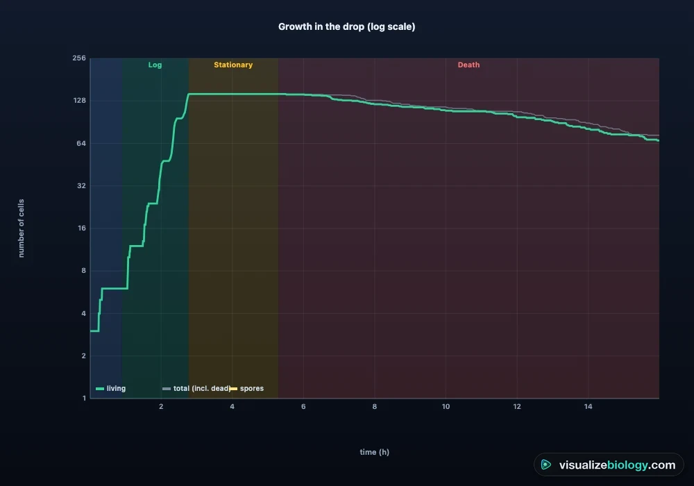
- Lag phase: the cells adjust to the new medium, making enzymes and ribosomes, but hardly divide.
- Log (exponential) phase: every cell divides once per generation time, so the number doubles at a steady rate: N = N₀ × 2ⁿ.
- Stationary phase: food runs out and waste builds up; cell division and death balance out. Spore formers make endospores now.
- Death (decline) phase: more cells die than are formed.
The generation time is g = t ÷ n, where n = 3.3 × log(N ÷ N₀) is the number of generations. It ranges from about 20 minutes for E. coli at 37 °C to 15–20 hours for Mycobacterium tuberculosis. Temperature, pH, oxygen and food all change it; the Graph tab shows each species' minimum, optimum and maximum temperature, from Listeria, which still grows in a refrigerator, to Thermus aquaticus, which grows best at 70 °C.
Respiration and fermentation in bacteria
Bacteria have no mitochondria. In aerobic respiration glycolysis and the Krebs cycle run in the cytoplasm and the electron transport chain sits in the plasma membrane, pumping protons out; the protons flowing back through ATP synthase make ATP (in theory up to 38 per glucose in a prokaryote). Some bacteria use nitrate or sulfate instead of oxygen (anaerobic respiration). Without an electron acceptor they ferment: pyruvate is turned into lactic acid, mixed acids or other products, which makes only 2–3 ATP per glucose. That is why a facultative anaerobe such as E. coli grows more slowly and to about a quarter of the cell density without oxygen. Bacteria are grouped by what oxygen does to them, and a thioglycolate tube shows the group at a glance:
| Group | Oxygen | Growth in thioglycolate | Example |
|---|---|---|---|
| Obligate aerobe | Needs it | Top only | Micrococcus, Mycobacterium, Pseudomonas |
| Facultative anaerobe | Uses it if present | All through, densest at the top | E. coli, Staphylococcus |
| Microaerophile | Needs a little (about 5 %) | A band just below the top | Helicobacter pylori |
| Aerotolerant anaerobe | Does not use it, not harmed | Even all through | Lactobacillus, Streptococcus |
| Obligate anaerobe | Killed by it | Bottom only | Clostridium |
Gene transfer: conjugation, transformation and transduction
Bacteria do not reproduce sexually, but they swap genes sideways (horizontal gene transfer). In conjugation a donor cell with a fertility (F) or resistance (R) plasmid builds a sex pilus, pulls a recipient close and passes it a copy of the plasmid. In transformation a competent cell takes up free DNA released by dead cells: in Griffith's 1928 experiment, harmless rough pneumococci took up capsule genes from heat-killed smooth ones and became deadly, and Avery showed in 1944 that the transforming substance is DNA. In transduction a bacteriophage carries a piece of DNA from one host to the next. All three can be watched in the simulator, and the pGLO lab uses transformation to put a jellyfish glow gene into E. coli.
Endospores
When food runs out, Bacillus and Clostridium make an endospore inside each cell in about eight hours: a dehydrated copy of the chromosome wrapped in a thick cortex and protein coats. Endospores survive boiling, drying, UV and disinfectants for years. Boiling for 10 minutes kills every growing cell but only a fraction of spores; an autoclave (steam at 121 °C for 15 minutes) kills them all, which is how labs and hospitals sterilize.
Antibiotics and antibiotic resistance
Antibiotics hit structures that bacteria have and human cells do not: penicillin and vancomycin block the building of peptidoglycan, so growing cells burst; tetracycline, erythromycin and gentamicin block 70S ribosomes; ciprofloxacin blocks DNA gyrase; rifampin blocks bacterial RNA polymerase; polymyxin breaks Gram-negative membranes. They do nothing against viruses. In a Kirby–Bauer test, disks of antibiotic on a lawn of bacteria leave clear zones of inhibition, wider when the bacterium is more susceptible. Resistance evolves by natural selection: resistant mutants or plasmid carriers already exist, and the antibiotic kills their susceptible neighbors. The evolution lab shows that a dose that kills susceptible cells but not resistant mutants (the mutant selection window) selects resistance fastest. This simulator is an educational model, not medical advice.
Experiments and course links
| Lab | What you find out | Course links |
|---|---|---|
| Gram stain three unknowns | Gram reaction, shape and arrangement; what each step does | AP Biology 2.1; college microbiology |
| The bacterial growth curve | Lag, log, stationary and death phases; generation time from OD₆₀₀ | OpenStax Microbiology 9.1; NGSS HS-LS2-1 |
| Serial dilution and plate count | CFU/mL from a countable plate (30–300 colonies) | Lab skills; college microbiology |
| Kirby–Bauer disk diffusion | Zones of inhibition; susceptible, intermediate or resistant | AP Biology 7.2; GCSE/A-level required practical |
| Thioglycolate tubes and catalase | The five oxygen classes | OpenStax Microbiology 9.3 |
| pGLO transformation | Gene expression with arabinose; transformation efficiency | AP Biology Investigation 8; Unit 6 |
| Evolution of antibiotic resistance | Natural selection and the mutant selection window | AP Biology 7.2–7.4; NGSS HS-LS4-4, HS-LS4-5 |
Who uses this virtual bacteria lab?
High school and college students use it to learn the parts of a bacterial cell, the types of bacteria and the Gram stain, and to practice growth-curve, generation-time, CFU and transformation-efficiency calculations with worked answers. Teachers project the 3D cell and the growth curve, and assign the labs when there are no cultures, incubators or biosafety facilities, or as a pre-lab before the real thing. It is an educational model built from published measurements, it needs no login, and it runs on school Chromebooks.
Frequently asked questions
What are the main parts of a bacterial cell?
Every bacterium has a plasma membrane, cytoplasm, 70S ribosomes and a nucleoid, the region where its single circular chromosome lies (there is no nucleus). Almost all have a peptidoglycan cell wall, and Gram-negative bacteria also have an outer membrane. Many have plasmids (small extra rings of DNA), and some have a capsule, pili for sticking and passing DNA, and flagella for swimming.
What is the difference between Gram-positive and Gram-negative bacteria?
Gram-positive bacteria have a thick cell wall of peptidoglycan (20 to 80 nm) with teichoic acids and no outer membrane, so they keep the crystal violet in a Gram stain and look purple. Gram-negative bacteria have a thin peptidoglycan layer (2 to 7 nm) inside an outer membrane covered in lipopolysaccharide; alcohol washes the crystal violet out, the safranin counterstain dyes them pink, and their outer membrane keeps out many antibiotics.
How do bacteria reproduce?
By binary fission, a form of asexual reproduction. The cell copies its circular chromosome starting from one origin, the two copies move apart as the cell grows, a ring of FtsZ protein pinches the cell in the middle and a septum of new wall divides it into two identical daughter cells. In good conditions E. coli divides every 20 minutes. Bacteria also gain new genes by conjugation, transformation and transduction, but these are not reproduction.
What are the four phases of bacterial growth?
In a closed culture: the lag phase, when cells adjust to the new medium and make enzymes but hardly divide; the log (exponential) phase, when the number doubles every generation time; the stationary phase, when food runs out and waste builds up so the number stays constant; and the death (decline) phase, when more cells die than divide. Plotted on a log scale, the log phase is a straight line.
How do antibiotics kill bacteria without harming human cells?
They target structures that bacteria have and our cells do not, or have in a different form: penicillins and vancomycin block peptidoglycan cell wall synthesis, tetracycline and erythromycin block 70S ribosomes, ciprofloxacin blocks DNA gyrase and rifampin bacterial RNA polymerase. Antibiotics do not work on viruses. Resistance evolves by natural selection when the drug kills susceptible cells and leaves resistant mutants or plasmid carriers to multiply.
Do bacteria need oxygen?
It depends on the species. Obligate aerobes (such as Mycobacterium tuberculosis) need oxygen for aerobic respiration; obligate anaerobes (such as Clostridium) are killed by it; facultative anaerobes (such as E. coli) respire with oxygen and ferment without it; microaerophiles (such as Helicobacter) need a little; aerotolerant anaerobes (such as Lactobacillus) ferment whether or not oxygen is present. A thioglycolate tube shows which type a bacterium is.
Explore related simulators
Compare a bacterium with the animal and plant cells of the Virtual Living Cell (prokaryote vs eukaryote), and with a single-celled eukaryote in the Virtual Amoeba Simulator, which eats bacteria by phagocytosis. The phages that infect bacteria, and the difference between a virus and a bacterium, are in the Virus Virtual Lab. Find Gram-stained bacteria on a slide with the Virtual Microscope, see the DNA a bacterium copies in the DNA structure 3D model, compare binary fission with the Mitosis Simulator, and see decomposers at work in Build the Food Chain. Meet the trillions of bacteria living in the colon in the Human Digestive System Simulator. See soil bacteria at work in an ecosystem, fixing nitrogen in root nodules, nitrifying and denitrifying, in the Nitrogen Cycle Simulator.
Sources: OpenStax Microbiology (2016), chapters 3, 8, 9, 11 and 14; Madigan et al., Brock Biology of Microorganisms; Rosso et al., Journal of Theoretical Biology 162 (1993), the cardinal temperature model; Baranyi and Roberts, International Journal of Food Microbiology 23 (1994), the lag phase; Cooper and Helmstetter, Journal of Molecular Biology 31 (1968), the E. coli cell cycle; Bauchop and Elsden, Journal of General Microbiology 23 (1960), growth yield and ATP; CLSI M02 and M100, disk diffusion quality-control ranges and breakpoints (simplified here for teaching); Bio-Rad, pGLO Bacterial Transformation Kit; College Board, AP Biology Course and Exam Description. Sizes, rates and zones are an educational model based on these sources.
Updated