
DNA Structure 3D Model
Watch the double helix being built from a single nucleotide, then take it apart: turn it, name every atom, pair the bases, unzip and melt it, and compare A-, B- and Z-DNA. Built atom by atom from real geometry, from high school to college.
Live math
Lab notebook
Every sequence you build, every mutation and every measurement is written here. Export a CSV, or turn the session into a PDF lab report.
| # | Sample | Setup | Result |
|---|
Concept library
Click "New problem" to begin.
Click "Start quiz" for 5 questions: the parts of a nucleotide, nomenclature, base pairing, the double helix and its stability.
1 Overview
Simulate builds a DNA double helix in front of you, from one nucleotide to the finished helix, and then hands it to you: turn it, name every part, change the sequence, unzip or melt it, and switch between A-, B- and Z-DNA. Beside the model, close-up views show the chemistry of a nucleotide, one base pair, both strands of the sequence, the three forms of the helix and an X-ray diffraction pattern. Experiment has four labs, ending in a PDF lab report. Quiz has a practice quiz with worked solutions and a 5-question quiz.
2 Getting started: the build
- The model opens in 3D on a computer or tablet and in 2D on a phone. The 2D | 3D switch changes it any time and remembers your choice.
- The build plays on its own in six chapters: a nucleotide → a strand → pairing → antiparallel strands → the twist → grooves. Press ▶ (or Space) to pause, Next ⏭ (or N) to step, or click a chapter on the strip. Skip ⇥ jumps to the finished helix; after that the same button builds it again.
- The caption under the model says what is happening. Changing the sequence, the form or unzipping ends the build and gives you the finished helix.
3 Turning, naming and selecting
- 3D: drag to turn, hold Ctrl (⌘ on a Mac) and scroll, or pinch, to zoom. A plain scroll always scrolls the page. On a phone turn it with two fingers.
- 2D: drag sideways to turn the helix; let go to let it coast.
- Point at any part to name it; click it to select that nucleotide. In ball & stick and space-filling you get each atom's name (C1′, O3′, N7…) and its role.
- The camera rail (Side, Axis, Groove, Close) frames the helix from the side, down its axis, into the major groove, or close on the selected nucleotide.
4 Model, sequence and helix controls
- Model: cartoon (backbone tubes and ring shapes), ball & stick or space-filling; color by part, base, strand or atom; hydrogens; slow spin; fade all but the selected nucleotide.
- Sequence: type strand 1 from 5′ to 3′ (4–30 bases) or pick a preset; the complementary strand is written for you. Select a pair and press A, T, G or C to mutate it: its partner changes with it.
- Helix: change form (A, B, Z), unzip the strands, or heat the DNA and watch A–T rich stretches open first.
- Show: 5′ → 3′ arrows, the grooves, and rulers for width, turn and rise.
5 The close-up views
- Nucleotide: the chemical structure of one nucleotide with carbons 1′–5′ numbered, its nucleoside and nucleotide names, and DNA vs RNA.
- Base pair: the pair drawn from its real atomic coordinates, hydrogen bonds with their lengths, the 1.08 nm C1′–C1′ width and which edge faces each groove.
- Sequence: both strands with their ends, composition (A = T, G = C), GC content and Tm. Click a letter to select its pair.
- A·B·Z: the three forms side by side, from the model. Click one to switch.
- X-ray: the fiber diffraction pattern computed from the helix: B-DNA gives Photo 51's X.
6 Experiment mode and the lab report
Four labs: Chargaff's rules from the base composition of eight organisms (including the single-stranded φX174), measuring the pitch and rise from Photo 51 by dragging lines onto it, melting five genomic DNAs in a heated spectrophotometer and plotting Tm against GC content, and building the model like Watson and Crick by testing which pairings fit. Each lab shows its aim, what you need and a safety note; the method ticks off as you go; a conclusion question checks your answer from your own data; and Create lab report builds a PDF.
7 SI and US units
Molecular sizes stay in nanometers (nm) and ångströms (1 Å = 0.1 nm) in both, because every biology and chemistry class measures molecules in them. US shows temperatures in °F (Tm 74 °C = 165 °F) and lengths you could hold in feet (the 2.2 m of DNA in one human cell is about 7.2 ft).
8 Keyboard
- Space plays or pauses the build, N goes to the next chapter, S opens Status.
- 1–5 switch the close-up view; C, B, F choose cartoon, ball & stick or space-filling; U unzips.
- With a pair selected, ← → move along the helix and Shift + A/T/G/C mutate it; Esc clears the selection.
- Right-click a view to save it as an image or copy the sequence.
DNA structure: an interactive 3D model of the double helix
DNA (deoxyribonucleic acid) carries the genetic instructions of every living thing. Its structure is famous because it explains how those instructions are stored and copied: two strands wound into a double helix, held together by pairs of bases that fit only one way. This free 3D DNA model builds that structure in front of you from a single nucleotide, then lets you take it apart, atom by atom, from the parts of a nucleotide to the grooves of the helix and the X-ray photograph that revealed it.
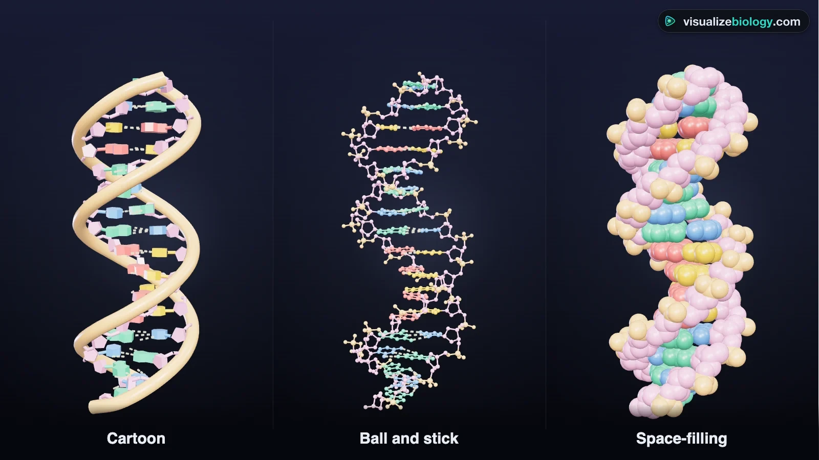
- Model
- Every atom from standard base geometry, with a sugar–phosphate backbone that closes with real bond lengths
- Views
- Cartoon, ball and stick, space-filling; nucleotide, base pair, sequence, A·B·Z and X-ray close-ups
- Change it
- Edit the sequence, mutate a pair, unzip, melt, switch between A-, B- and Z-DNA
- Labs
- Chargaff's rules, Photo 51, DNA melting and GC content, Watson–Crick model building
- Modes
- Simulate, Experiment (4 labs) and Quiz
- Runs on
- Any modern browser, including Chromebooks, iPads and phones. 3D on larger screens, 2D on phones.
- Price
- Free, with no account or download
What is DNA made of? The parts of a nucleotide
DNA is a polymer: a long chain of units called nucleotides. Each nucleotide has three parts: a phosphate group, a five-carbon sugar called deoxyribose, and a nitrogenous base. There are four bases: adenine (A), guanine (G), cytosine (C) and thymine (T). Adenine and guanine are purines, with two rings; cytosine and thymine are pyrimidines, with one.
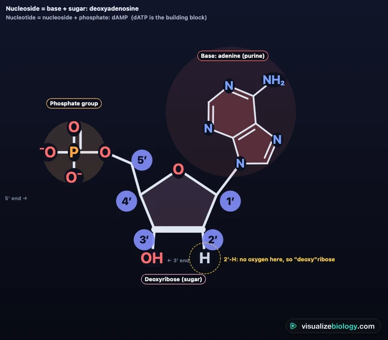
Nucleotide nomenclature: 1′ to 5′, nucleosides and nucleotides
The sugar's carbons are numbered with a prime, 1′ to 5′ ("one prime" to "five prime"), to tell them from the atoms of the base. The base is joined to carbon 1′ by an N-glycosidic bond, the phosphate sits on carbon 5′, and carbon 3′ carries the –OH that links to the next nucleotide. A base joined to the sugar is a nucleoside; add a phosphate and it becomes a nucleotide.
| Base | Type | DNA nucleoside | DNA nucleotide | In RNA |
|---|---|---|---|---|
| Adenine (A) | Purine | Deoxyadenosine | dAMP (built from dATP) | Adenosine, AMP |
| Guanine (G) | Purine | Deoxyguanosine | dGMP | Guanosine, GMP |
| Cytosine (C) | Pyrimidine | Deoxycytidine | dCMP | Cytidine, CMP |
| Thymine (T) | Pyrimidine | Thymidine | dTMP | Replaced by uracil: uridine, UMP |
Base pairing rules and Chargaff's rule
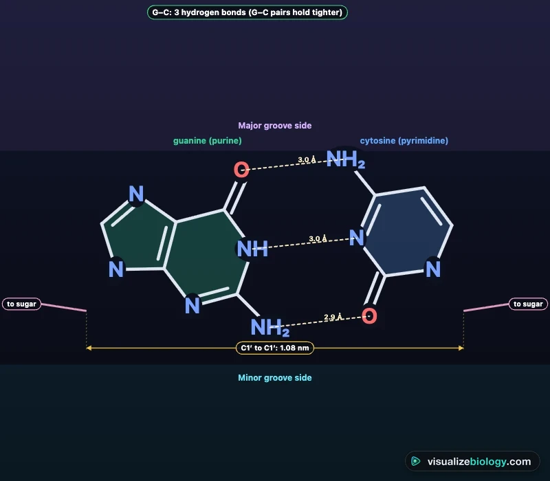
In the double helix, adenine always pairs with thymine and guanine with cytosine. An A–T pair is held by two hydrogen bonds and a G–C pair by three. Each pair is a purine opposite a pyrimidine, so every rung of the ladder is the same width and any sequence fits the same smooth backbone. This is the reason for Chargaff's rule: in double-stranded DNA the amount of A equals T and G equals C. The ratio of A + T to G + C differs between species, about 41 % G + C in humans and 51 % in E. coli.
Antiparallel strands: the 5′ and 3′ ends
Nucleotides join by phosphodiester bonds: the phosphate on one sugar's 5′ carbon bonds to the 3′ carbon of the next. A strand therefore has a direction, from a 5′ end (free phosphate) to a 3′ end (free –OH). The two strands of the helix run in opposite directions: they are antiparallel. Sequences are written 5′ → 3′, so the complementary strand of 5′-ATGCTA-3′ is 5′-TAGCAT-3′: pair each base, then reverse.
The double helix: dimensions and grooves
The sugar–phosphate backbones form the outside of the helix and the bases stack inside, 0.34 nm apart, like a spiral staircase. In B-DNA the helix is right-handed, 2 nm in diameter, with about 10.5 base pairs per turn. Because the two sugars of each pair lie on one side of it, the backbones are not evenly spaced: a wide major groove and a narrow minor groove wind round the helix. The base edges are most exposed in the major groove, where proteins such as transcription factors read the sequence without opening the helix.
length of B-DNA ≈ number of base pairs × 0.34 nm
A-DNA, B-DNA and Z-DNA
| A-DNA | B-DNA | Z-DNA | |
|---|---|---|---|
| Handedness | Right | Right | Left |
| Base pairs per turn | 11 | 10.5 | 12 |
| Rise per base pair | 0.26 nm | 0.34 nm | 0.37 nm |
| Diameter | 2.3 nm | 2.0 nm | 1.8 nm |
| Grooves | Narrow, deep major; wide, shallow minor | Wide major, narrow minor | Flat major, deep narrow minor |
| Where | Dehydrated DNA, RNA duplexes, DNA–RNA hybrids | Most DNA in cells | Alternating CG sequences, high salt, supercoiling |
How the structure of DNA was discovered
In 1950 Erwin Chargaff showed that A ≈ T and G ≈ C. In 1952 Rosalind Franklin and Raymond Gosling took Photo 51, an X-ray diffraction photograph of B-DNA fibers: its X shape revealed a helix, its layer lines a repeat of 3.4 nm, and its strong arcs the 0.34 nm stacking of the bases. In 1953 James Watson and Francis Crick built the double helix that fitted both. Watson, Crick and Maurice Wilkins shared the 1962 Nobel Prize; Franklin had died in 1958. The X-ray view of this model computes a fiber pattern from the helix, so you can measure it yourself.
What holds the double helix together?
Hydrogen bonds make base pairing specific, but most of the helix's stability comes from base stacking, the attraction between flat bases piled on top of each other. Heating breaks both, and the strands separate: this is DNA melting, measured as a 37 % rise in absorbance at 260 nm. The melting temperature (Tm) rises with GC content; for genomic DNA in SSC buffer, Tm = 69.3 + 0.41 × (%GC) °C.
DNA vs RNA
RNA uses ribose, which has an –OH on carbon 2′, and uracil instead of thymine, and it is usually single-stranded. Switch the nucleotide view to RNA to see both differences.
Who uses this DNA model?
High school and AP Biology students use it to learn the parts of DNA, the base pairing rules and antiparallel strands; college students use the atom names, the A, B and Z forms, melting thermodynamics and diffraction. Teachers project the build, pause on each chapter, and run the labs without equipment. It is an educational model built from published structural data, it needs no login, and it runs on school Chromebooks.
Frequently asked questions
What is the structure of DNA?
DNA is a double helix: two strands of nucleotides wound around each other like a twisted ladder. The sides of the ladder are sugar–phosphate backbones, and the rungs are pairs of bases held together by hydrogen bonds: adenine with thymine and guanine with cytosine. The two strands run in opposite directions (antiparallel). In the common B form the helix is right-handed, 2 nm wide, with a base pair every 0.34 nm and about 10.5 base pairs per turn.
What are the three parts of a nucleotide?
A phosphate group, a five-carbon sugar and a nitrogenous base. In DNA the sugar is deoxyribose and the base is adenine (A), thymine (T), guanine (G) or cytosine (C). The sugar's carbons are numbered 1′ to 5′: the base is attached to carbon 1′ and the phosphate to carbon 5′, and carbon 3′ links to the next nucleotide's phosphate.
What are the base pairing rules in DNA?
Adenine pairs with thymine (A–T) by two hydrogen bonds, and guanine pairs with cytosine (G–C) by three. Each pair is a purine (A or G, two rings) with a pyrimidine (T or C, one ring), so every rung of the helix has the same width. That is why, in double-stranded DNA, the amount of A equals the amount of T and G equals C (Chargaff's rule).
What does antiparallel mean, and what are the 5′ and 3′ ends of DNA?
Each strand has a direction set by its sugars: the 5′ end has a free phosphate on carbon 5′, and the 3′ end a free –OH on carbon 3′. In the double helix the two strands point opposite ways: one runs 5′ to 3′ and its partner 3′ to 5′. Sequences are written 5′ to 3′, and DNA polymerase can only add nucleotides to a 3′ end.
What is the difference between the major and minor groove of DNA?
Because the two sugars of a base pair sit on one side of it, the backbones are not evenly spaced around the helix. The wide, deep gap is the major groove (about 1.2 nm wide in B-DNA) and the narrow one is the minor groove (about 0.6 nm). The edges of the bases are more exposed in the major groove, so most proteins that read DNA, such as transcription factors, bind there.
What is the difference between A-DNA, B-DNA and Z-DNA?
B-DNA is the usual form in cells: right-handed, about 10.5 base pairs per turn and 2 nm wide. A-DNA is right-handed but shorter and wider, with 11 base pairs per turn and tilted base pairs; dehydrated DNA and double-stranded RNA take this form. Z-DNA is left-handed, with 12 base pairs per turn and a zigzag backbone; it forms in alternating purine–pyrimidine sequences such as CGCGCG.
Explore related simulators
See how DNA is copied and shared out when cells divide in the Mitosis Simulator, follow chromosomes and crossing over into gametes with the Meiosis Simulator, and look at nuclei and chromosomes under the Virtual Microscope. See DNA (and RNA) packed inside viruses, and HIV copying RNA back into DNA, in the Virus Virtual Lab. Read what the bases spell, three at a time, with the Interactive Codon Chart and DNA to Protein Translator: paste any sequence and watch it become a protein. Follow alleles, the versions of a gene, from parents to offspring with the Interactive Punnett Square Calculator.
Sources: OpenStax Biology 2e, chapter 14; Watson & Crick and Franklin & Gosling, Nature 171 (1953); Olson et al., J. Mol. Biol. 313 (2001), standard base geometry; Saenger, Principles of Nucleic Acid Structure (1984), helix parameters; SantaLucia, PNAS 95 (1998), nearest-neighbor thermodynamics; Marmur & Doty, J. Mol. Biol. 5 (1962), Tm and GC content; Chargaff, Experientia 6 (1950). The Z-DNA backbone is a schematic fiber model. This is an educational model.
Updated