
Meiosis Simulator
Follow one cell through meiosis I and II in 3D: homologs pair up, swap pieces and separate, and four genetically different gametes come out, each with its own genotype. Then score Sordaria asci to map a gene, as in AP Biology Lab 7.
How to score a hybrid ascus
Each ascus holds 8 spores in the order the nuclei divided: meiosis I, meiosis II, then one mitosis.
map units = (crossover asci ÷ total hybrid asci × 100) ÷ 2
Click an ascus on the slide, or hover over it and press 4, C or X. Drag the slide to find more rosettes.
Live math
Lab notebook
Every meiosis you finish, every set of gametes you record and every count you make is written here. Export a CSV, or turn the session into a PDF lab report.
| # | Sample | Setup | Result |
|---|
Concept library
Click "New problem" to begin.
Click "Start quiz" for 5 questions: naming the stage, chromosome counts, genetic variation, the Sordaria lab and mitosis vs meiosis.
1 Overview
Simulate follows one diploid germ cell through interphase, meiosis I and meiosis II until four haploid gametes come out. Every chromatid is colored by the parent it came from (pink from the mother, blue from the father) and carries alleles, so you can see exactly which mix each gamete gets. Beside the cell you can watch the gametes and their genotypes, a close-up of one homologous pair, graphs of DNA and chromosome number, and mitosis and meiosis side by side. Experiment has five labs, from scoring Sordaria asci to a chi-square test, ending in a PDF lab report. Quiz has a practice quiz with worked solutions and a 5-question quiz.
2 Getting started
- The cell opens in 3D on a computer or tablet and in 2D on a phone. The 2D | 3D switch changes it any time and remembers your choice.
- Press ▶ (or Space) to play. Teach gives every phase time on screen; Real keeps the true proportions, so you see that prophase I takes most of the time.
- Next phase ⏭ (or N) jumps to the start of the next phase and pauses. Click any segment of the phase strip or the graph to jump straight there.
- The caption says what is happening, and the arrows show where things move: homologs pairing, chromosomes to the plate, homologs and then chromatids to the poles.
- Click a part of the cell (a chromatid, the spindle, a centrosome, a polar body) to name it and read what it does.
3 The cell and genetic variation
- Sperm or egg: a spermatocyte divides equally into four sperm cells. An oocyte divides unequally twice, giving one large egg and three small polar bodies, and its spindles have no centrosomes.
- 2n = 4, 6 or 8 keeps the picture clear. Each pair carries one gene (A, B, D, E) with a dominant allele from one parent and a recessive one from the other.
- Crossing over swaps matching pieces of non-sister chromatids in pachytene; switch it off to see assortment alone. ✕ New crossovers puts them somewhere else.
- Pair ⇄ buttons flip how a pair faces at metaphase I (independent assortment); 🎲 Random line-up flips them all at random. Both work until anaphase I.
- Errors: make pair 1 fail to separate in meiosis I or in meiosis II, and compare the gametes.
4 Gametes, Pair, Graph and vs Mitosis views
- Gametes: the four products with their chromosomes and genotypes, and every combination the cell could make, with the ones it made lit.
- Pair: one homologous pair close up: synapsis, the synaptonemal complex, the chiasma and which chromatids crossed over.
- Graph: DNA per cell (2C → 4C → 2C → 1C), chromosomes per cell, number of cells, or the tally of many gametes.
- vs Mitosis: the two divisions side by side at the same moment, with a table of the differences.
5 The Sordaria lab
Drag the slide to a rosette of asci and click an ascus (or hover and press 4, C or X) to call it 4:4 (no crossover), crossover (2:2:2:2 or 2:4:2) or not countable (all one color, or pale and unripe). The tally and the map distance update as you go. Check my calls marks each of your calls green or red against an expert's.
6 Experiment mode and the lab report
Five labs: crossing over in Sordaria (AP Lab 7 Part 4), independent assortment with chi-square, linked genes and recombination frequency, nondisjunction in meiosis I or II, and predicting the gametes (AP Lab 7 Part 3). Each shows its aim, what you need and a safety note. The method ticks off as you go, a conclusion question checks your answer from your own data, and Create lab report builds a PDF with your results, graph, worked calculations and sources of error.
7 Quiz
The practice quiz asks you to name the stage from a drawing, count chromosomes, chromatids, tetrads and DNA, work out gamete combinations, map distances and chi-square, and explain nondisjunction and the differences between mitosis and meiosis, with a worked solution for each. Take the quiz gives five mixed questions and a star rating.
8 Keyboard and gestures
- Space plays or pauses, N goes to the next phase, S opens Status, and E, P, G, V switch to gamEtes, Pair, Graph or vs mitosis.
- On the Sordaria slide, hover over an ascus and press 4 (4:4), C (crossover) or X (not counted).
- Scrolling over the 3D cell always scrolls the page. To zoom, hold Ctrl (⌘ on a Mac) and scroll, or pinch on a trackpad. On a phone, one finger scrolls the page; turn the 3D cell with two fingers.
- Right-click a view to save it as an image or copy your results.
Stages of meiosis: an interactive meiosis simulator and Sordaria lab
Meiosis is the cell division that makes gametes: sperm and eggs. One diploid cell divides twice and gives four haploid cells, each with one chromosome from every pair, and no two of them alike. That is why you are not a copy of either parent, and why brothers and sisters differ. This free meiosis simulator lets you watch it happen in 3D, follow one pair of chromosomes from start to finish, and see which alleles each gamete gets. Then, in the virtual lab, you score Sordaria asci to measure crossing over yourself, as in AP Biology Lab 7.
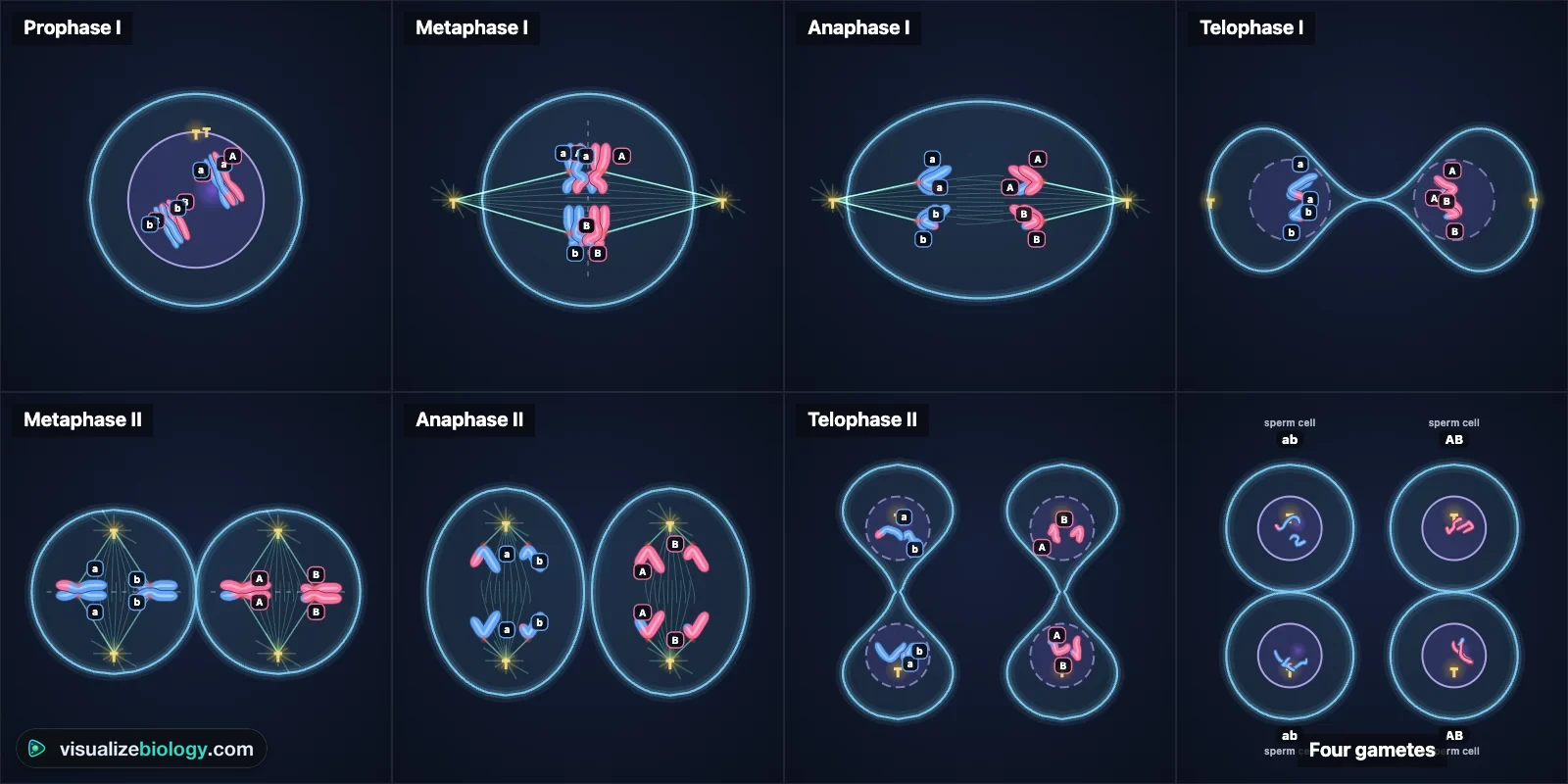
- Cells
- A spermatocyte (four sperm) or an oocyte (one egg and three polar bodies), with 2n = 4, 6 or 8
- Stages
- G1, S, G2; prophase I (leptotene to diakinesis), metaphase I, anaphase I, telophase I; interkinesis; prophase II to telophase II
- Variation
- Crossing over, independent assortment, linked genes, nondisjunction in meiosis I or II
- Labs
- Sordaria map units, chi-square test of assortment, recombination frequency, nondisjunction, predicting gametes
- Modes
- Simulate, Experiment (5 labs) and Quiz
- Runs on
- Any modern browser, including Chromebooks, iPads and phones. 3D on larger screens, 2D on phones.
- Price
- Free, with no account or download
What is meiosis?
Meiosis is a reduction division: it halves the number of chromosomes. A diploid (2n) cell has two sets of chromosomes, one from each parent; in humans that is 23 pairs, or 46. Each gamete must be haploid (n = 23) so that when a sperm fertilizes an egg the zygote is diploid again. Before meiosis the cell copies its DNA once, in S phase, so each chromosome is two sister chromatids. It then divides twice: meiosis I separates the homologous chromosomes, and meiosis II separates the sister chromatids. The result is four haploid cells.
The stages of meiosis I and meiosis II
| Stage | What happens | Chromosomes per cell (human) |
|---|---|---|
| Prophase I | Chromosomes condense and homologs pair up (synapsis), held by the synaptonemal complex. Non-sister chromatids cross over. The nuclear envelope breaks down. | 46, as 23 tetrads |
| Metaphase I | Homologous pairs line up on the metaphase plate, each facing a random way. | 46 |
| Anaphase I | Homologs separate to opposite poles. Sister chromatids stay together. | 46 |
| Telophase I and cytokinesis | Two haploid cells, each chromosome still two chromatids. | 23 |
| Prophase II | A new spindle forms in each cell. No DNA is copied. | 23 |
| Metaphase II | Single chromosomes line up on the plate, as in mitosis. | 23 |
| Anaphase II | Sister chromatids separate to opposite poles. | 46 for a moment |
| Telophase II and cytokinesis | Four haploid cells, genetically different. | 23 |
Prophase I is by far the longest stage. Biologists split it into five: leptotene (threads appear), zygotene (homologs zip together), pachytene (fully paired; crossing over happens), diplotene (homologs pull apart except at the chiasmata) and diakinesis (fully condensed). In a human spermatocyte, meiosis I takes about 24 days, nearly all of it prophase I.
How meiosis makes genetic variation
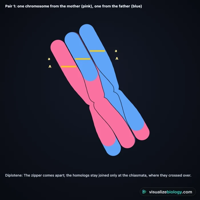
Crossing over swaps matching pieces between a maternal and a paternal chromatid of the same pair, making chromosomes that never existed in either parent. Independent assortment means each pair lines up at metaphase I independently of the others, so a cell with n pairs can make 2n kinds of gamete: about 8.4 million in humans before any crossing over. Random fertilization then combines one of millions of sperm with one of millions of eggs, giving over 70 trillion possible zygotes. Together these are the new genetic combinations through meiosis in NGSS HS-LS3-2.
Mitosis vs meiosis
Both divisions use the same spindle and the same stage names, which is why they are easy to mix up. The difference is what separates and how many times. Use the vs Mitosis view in the simulator to watch both at the same moment.
| Mitosis | Meiosis | |
|---|---|---|
| Purpose | Growth, repair, asexual reproduction | Gametes for sexual reproduction |
| Divisions | One | Two (meiosis I and II) |
| Daughter cells | Two, diploid (2n) | Four, haploid (n) |
| Genetically | Identical to the parent and each other | Different from the parent and each other |
| Homologs pair (synapsis) | No | Yes, in prophase I |
| Crossing over | No | Yes, in prophase I |
| At metaphase | Single chromosomes on the plate | Pairs (meiosis I), then single chromosomes (meiosis II) |
| At anaphase | Sister chromatids separate | Homologs separate (I), then sister chromatids (II) |
| Where in the body | Almost all body cells | Only in testes and ovaries (anthers and ovules in plants) |
To watch mitosis on its own, stage by stage, with an onion root tip lab, use the Mitosis Simulator.
Spermatogenesis and oogenesis
In the testes, each primary spermatocyte divides equally to give four spermatids, which mature into sperm. In the ovaries, both divisions are unequal: nearly all the cytoplasm stays in one cell, so a primary oocyte gives one egg and three polar bodies (often only two, as the first polar body may not divide), which break down. Human oocytes start meiosis before birth, pause in prophase I until puberty, and pause again at metaphase II, finishing meiosis II only if a sperm arrives. Switch the simulator to an oocyte to see both unequal divisions.
Nondisjunction
If a pair of homologs fails to separate in meiosis I, all four gametes have the wrong number of chromosomes: two n + 1 and two n − 1. If sister chromatids fail to separate in meiosis II, only one of the two cells is affected: one n + 1, one n − 1 and two normal gametes. A gamete with an extra chromosome fertilized by a normal one gives a trisomy (2n + 1), such as Down syndrome (trisomy 21); one with a missing chromosome gives a monosomy, such as Turner syndrome (45,X). The risk rises with the mother's age, because her eggs have waited in prophase I for decades. This is an educational model, not medical advice.
The Sordaria lab: mapping a gene with crossing over
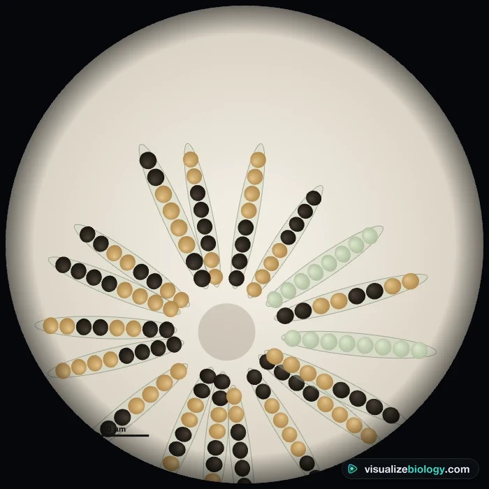
In the fungus Sordaria fimicola, each ascus holds eight spores in a row, in the order the nuclei divided: meiosis followed by one mitosis. When a black and a tan strain meet, the hybrid asci show whether a crossover happened between the spore-color gene and its centromere. No crossover gives a 4:4 pattern; a crossover gives 2:2:2:2 or 2:4:2.
map distance = (crossover asci ÷ total hybrid asci × 100) ÷ 2
Worked example. A student scores 120 hybrid asci: 64 are 4:4 and 56 show a crossover. Percent crossover asci = 56 ÷ 120 × 100 = 46.7 %. Divide by 2, because a crossover involves only two of the four chromatids, so only half the spores in a crossover ascus are recombinant: the gene is about 23 map units from its centromere. The published value is about 26.
Experiments and course links
| Lab | What you find out | Course links |
|---|---|---|
| Crossing over in Sordaria | Map distance of the spore-color gene from its centromere | AP Biology Lab 7 Part 4 |
| Independent assortment | Do AaBb gametes fit 1:1:1:1? A χ² goodness-of-fit test | AP Unit 5, NGSS HS-LS3-2, A-level |
| Linked genes | Recombination frequency and map distance between two genes | AP Unit 5, A-level, IB D3.2 |
| Nondisjunction | Meiosis I vs meiosis II errors and the gametes they make | AP Unit 5, NGSS HS-LS3-2 |
| Predict the gametes | Model meiosis and test your prediction | AP Biology Lab 7 Part 3, GCSE |
Who uses this meiosis simulator?
Students use it to learn the stages of meiosis 1 and 2, to see where variation comes from, and to practice chromosome counts, map units and chi-square with worked answers. Teachers project it and pause on prophase I, flip a pair to show independent assortment, and run the Sordaria lab when there are no cultures to grow. It is an educational model built from textbook values, it needs no login, and it runs on school Chromebooks.
Frequently asked questions
What are the stages of meiosis?
Meiosis has two divisions. Meiosis I: prophase I (homologs pair and cross over), metaphase I (pairs line up on the plate), anaphase I (homologs separate) and telophase I. Meiosis II: prophase II, metaphase II (single chromosomes line up), anaphase II (sister chromatids separate) and telophase II. The DNA is copied once, in S phase before meiosis I, and not again before meiosis II.
What is the difference between mitosis and meiosis?
Mitosis is one division that makes two diploid cells identical to the parent, for growth and repair. Meiosis is two divisions that make four haploid cells, all genetically different, for sexual reproduction. Only in meiosis do homologous chromosomes pair up, cross over and separate (in meiosis I); meiosis II separates sister chromatids as mitosis does.
What happens during crossing over?
In prophase I, when homologous chromosomes are paired as a tetrad, a non-sister chromatid from each homolog breaks at the same point and the two rejoin to each other, swapping matching pieces. The crossover holds the pair together as a chiasma until anaphase I. Alleles that were on the same chromosome can end up apart, so each chromatid becomes a new mix of the mother's and the father's genes.
What is independent assortment?
At metaphase I each pair of homologs lines up with its maternal or its paternal chromosome facing a given pole at random, independently of the other pairs. A cell with n pairs can therefore make 2 to the power n kinds of gamete from assortment alone: 2^23, about 8.4 million, in humans. It is why genes on different chromosomes are inherited independently, as in Mendel's second law.
How do you calculate map units in the Sordaria lab?
Count hybrid asci only: 4:4 asci had no crossover between the spore-color gene and its centromere; 2:2:2:2 and 2:4:2 asci had one. Map distance = (crossover asci ÷ total hybrid asci × 100) ÷ 2. You divide by 2 because a crossover involves only two of the four chromatids, so only half of the spores in a crossover ascus are recombinant. The published distance is about 26 map units.
What is nondisjunction?
Nondisjunction is a failure of chromosomes to separate. In meiosis I a pair of homologs goes to the same cell, so all four gametes have the wrong number: two n + 1 and two n − 1. In meiosis II a pair of sister chromatids fails to separate in one cell, giving one n + 1, one n − 1 and two normal gametes. An n + 1 gamete fertilized by a normal one gives a trisomy, such as Down syndrome (trisomy 21).
Explore related simulators
Watch body cells divide in the Mitosis Simulator, look at real cells and their nuclei with the Virtual Microscope, see how a heart works in the Heart & Cardiac Cycle simulator, and zoom into the molecule every chromosome is made of with the DNA structure 3D model. Cross the gametes meiosis makes, one, two or three genes at a time, and predict the offspring with the Interactive Punnett Square Calculator. See where a plant's gametes meet, pollen on the stigma and the egg in the ovule, and watch double fertilization in the Pollination Simulator.
Sources: OpenStax Biology 2e, chapter 11 Meiosis and Sexual Reproduction; Alberts et al., Molecular Biology of the Cell, 6th ed., chapter 17 (meiosis); Olive, American Journal of Botany 43 (1956), on spore-color mutants of Sordaria fimicola; College Board, AP Biology Investigative Labs, Investigation 7. Stage durations are an educational model based on human spermatogenesis (about 64–74 days in all, meiosis I about 24 days).
Updated