
Interactive Codon Chart and DNA to Protein Translator
Read the genetic code on a wheel or a table, then paste any DNA or mRNA and watch a ribosome translate it codon by codon. Change one base and see whether the mutation is silent, missense, nonsense or a frameshift.
Open Mutate and tap any base of the DNA to change it, add a base after it or delete it.
Your protein
Lab notebook
Every sequence you translate and every mutation you make is written here with its result. Export a CSV, or turn the session into a PDF lab report.
| # | Gene | Change | Result | Protein |
|---|
Concept library
Click "New problem" to begin.
Click "Start quiz" for 5 questions on reading the codon chart, DNA to mRNA, translation, anticodons and mutations.
1 Overview
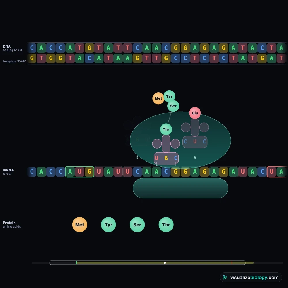
Simulate shows the genetic code as a codon chart on the left and a translator on the right. Pick a gene in the top bar (or type your own sequence in the Sequence panel), press ▶ and watch the DNA transcribed into mRNA and the mRNA translated into protein, with each codon lit up on the chart as the ribosome reads it. Experiment has six labs that end in a PDF lab report, and Quiz has a practice quiz with worked solutions and a 5-question quiz.
2 Reading the codon chart
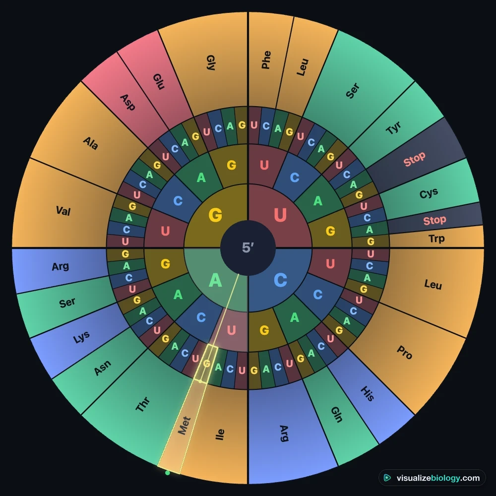
- Wheel: find the codon's first base in the centre ring, its second base in the next ring out and its third base in the third ring; the amino acid is on the rim. Table: first base on the left, second base along the top, third base on the right.
- Hover over or tap any codon to see its amino acid (one- and three-letter code), its anticodon and its DNA triplets. Tap an amino acid to light up every codon for it, or type one into Find an amino acid.
- The Codon panel lists the nine codons one base away and says whether each change would be silent, missense or a stop. Colour the chart by amino-acid class, hydropathy or the number of codons.
3 Translating a sequence
- Choose what you are typing: DNA coding strand (5′ → 3′), DNA template strand (written 3′ → 5′ under the coding strand, or 5′ → 3′ as databases print it) or mRNA. Spaces, numbers and a FASTA header are ignored; letters that are not bases are reported.
- Press Translate. The chips under the translator choose where reading starts: at the first AUG, as a ribosome does, or from the first, second or third base (frame 1, 2 or 3).
- ▶ plays transcription and translation; › steps one codon; the speed can be Teach, Fast or Real (a human ribosome reads about 6 codons a second). Your protein shows the result in one- and three-letter code, ready to copy.
4 Mutations
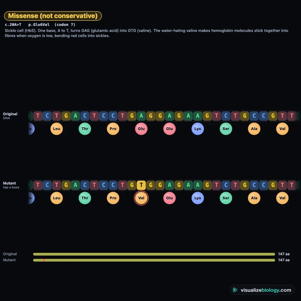
Open Mutate and tap a base of the DNA to change it, insert a base after it or delete it. The original and the mutant are shown one above the other, with the first changed amino acid marked and the kind of mutation named: silent, missense (conservative or not), nonsense, frameshift, in-frame insertion or deletion, start lost or stop lost. Real mutations loads sickle cell, hemoglobin C, two β-thalassemias and cystic fibrosis ΔF508. Undo with ↶ or Z.
5 Frames and the protein
Frames translates all six reading frames (three on each strand) and draws the open reading frames (AUG to a stop) as bars; tap one to translate it. Protein draws the chain coloured by amino-acid class, with its length, mass, net charge and the share of each class.
6 Experiment mode and the lab report
Six labs: decode a hidden message, DNA to protein by hand, the sickle cell mutation, types of mutation, which codon position is safest to change, and find the gene (open reading frames). Each shows its aim, what you need and a real-lab note; the method ticks off as you work, a conclusion question checks your result, and Create lab report builds a PDF with your gene, results and working.
7 Quiz and units
The practice quiz covers reading the chart, DNA to mRNA, mRNA to protein, codons and anticodons, counting bases, codons and amino acids, mutations and the properties of the code, each with a worked solution. Take the quiz gives five mixed questions and a star rating. The SI (m) | US (ft) switch changes everyday lengths (the 2 m of DNA in one cell is 6.6 ft); molecules stay in nanometers and bases, as in every biology class.
8 Keyboard and gestures
- Space plays or pauses, → steps one codon, W and T switch between wheel and table, Z undoes a mutation, S opens Status and Esc clears the selection.
- Drag the translator sideways to scroll along a long gene. A plain scroll over the chart or the translator scrolls the page.
- Right-click the chart or the translator to save it as an image or copy the sequence and protein.
The codon chart: how to read the genetic code
Every protein in your body is built from a recipe written in DNA, four letters long. The genetic code is the dictionary that turns that recipe into a protein: each group of three bases in messenger RNA, a codon, stands for one of twenty amino acids or for "stop". There are 4 × 4 × 4 = 64 codons, and a codon chart lists them all. This interactive codon chart lets you read the code as a wheel or as the classic table, and then use it: paste any DNA or mRNA and watch it translated, codon by codon, into a protein.
- Charts
- Codon wheel and the classic 4 × 4 codon table, coloured by class, hydropathy or number of codons
- Translator
- DNA coding or template strand, or mRNA (FASTA accepted), from the first AUG or any of six reading frames
- Mutations
- Silent, missense, nonsense, frameshift, in-frame deletions; sickle cell, β-thalassemia, ΔF508
- Modes
- Simulate (with animated translation), Experiment (6 labs) and Quiz
- Price
- Free, with no account or download; works on Chromebooks, tablets and phones
How to read a codon chart
Always work from the mRNA codon, read 5′ to 3′. On the table, find the first base in the left-hand column, move across to the column of the second base, and pick the line of the third base from the right-hand column. On the wheel, start at the centre with the first base and move outwards through the second and third rings to the amino acid on the rim. Try UGG: U in the centre, G next, G last, and you reach tryptophan. If you are given DNA, turn it into mRNA first (below).
| 1st base | 2nd: U | 2nd: C | 2nd: A | 2nd: G | 3rd base |
|---|---|---|---|---|---|
| U | UUU Phe | UCU Ser | UAU Tyr | UGU Cys | U |
| UUC Phe | UCC Ser | UAC Tyr | UGC Cys | C | |
| UUA Leu | UCA Ser | UAA Stop | UGA Stop | A | |
| UUG Leu | UCG Ser | UAG Stop | UGG Trp | G | |
| C | CUU Leu | CCU Pro | CAU His | CGU Arg | U |
| CUC Leu | CCC Pro | CAC His | CGC Arg | C | |
| CUA Leu | CCA Pro | CAA Gln | CGA Arg | A | |
| CUG Leu | CCG Pro | CAG Gln | CGG Arg | G | |
| A | AUU Ile | ACU Thr | AAU Asn | AGU Ser | U |
| AUC Ile | ACC Thr | AAC Asn | AGC Ser | C | |
| AUA Ile | ACA Thr | AAA Lys | AGA Arg | A | |
| AUG Met (start) | ACG Thr | AAG Lys | AGG Arg | G | |
| G | GUU Val | GCU Ala | GAU Asp | GGU Gly | U |
| GUC Val | GCC Ala | GAC Asp | GGC Gly | C | |
| GUA Val | GCA Ala | GAA Glu | GGA Gly | A | |
| GUG Val | GCG Ala | GAG Glu | GGG Gly | G |
From DNA to mRNA to protein
A gene has two strands. RNA polymerase reads the template strand and builds mRNA that pairs with it: A with U, T with A, G with C and C with G. So the mRNA has the same sequence as the other strand, the coding strand, with U in place of T. The template TAC CGA ATT becomes the mRNA AUG GCU UAA; the coding strand ATG GCT TAA gives the same mRNA. In translation, a ribosome moves along the mRNA one codon at a time. Each transfer RNA carries one amino acid and has an anticodon that pairs with its codon (UAC pairs with AUG), and the ribosome joins the amino acids into a chain. A human ribosome adds about six amino acids a second.
Start and stop codons
Translation begins at the first AUG, which codes for methionine and fixes the reading frame: from there the mRNA is read in non-overlapping threes. It ends at one of the three stop codons, UAA, UAG or UGA, which no tRNA matches; a release factor binds and the protein is let go. A stretch from AUG to a stop is an open reading frame. Read the same mRNA from one base later and every codon changes, which is why an insertion or deletion can ruin a whole protein.
Types of point mutations
| Mutation | What changes | Effect on the protein | Real example |
|---|---|---|---|
| Silent | One base, but the codon still codes for the same amino acid | None | GCU → GCC (both alanine) |
| Missense | One base, and the codon codes for a different amino acid | One amino acid changed | Sickle cell: GAG → GUG, Glu6Val in β-globin |
| Nonsense | One base turns a codon into a stop codon | Protein cut short | β-thalassemia: CAG → UAG at codon 39 |
| Frameshift | Bases inserted or deleted, not in a multiple of three | Every amino acid after it changes; usually an early stop | β-thalassemia codons 41/42 (−TTCT) |
| In-frame deletion | Three bases (one codon) lost | One amino acid missing | Cystic fibrosis ΔF508 |
The code is degenerate: most amino acids have more than one codon, and the extra codons usually differ only in the third base. Of the 549 possible single-base changes of the 61 sense codons, 134 are silent, and 126 of those are at the third base. No change at the second base is ever silent. That is why the third position is said to wobble, and why a tRNA can often read more than one codon.
Experiments and course links
| Lab | What you find out | Course links |
|---|---|---|
| Decode a hidden message | How to read the codon chart, start and stop | NGSS HS-LS1-1; GCSE 4.6.1.5 |
| DNA to protein by hand | Template strand → mRNA → anticodons → protein | AP Biology 6.3–6.4; A-level 3.4.2 |
| The sickle cell mutation | How one base changes a protein and a person | HS-LS3-2; AP Biology 6.7 |
| Types of mutation | Silent, missense, nonsense and frameshift side by side | GCSE 4.6.1.7; IB D1.3 |
| Which base is safest to change? | Degeneracy and the wobble position, from your own data | A-level 3.8.1; AP Biology 6.4 |
| Find the gene | Reading frames and open reading frames on both strands | AP Biology 6.3; IB D1.2 |
Who uses this codon chart?
Students use it to look up codons, to check their DNA-to-mRNA and mRNA-to-protein answers, and to practise mutation questions with worked solutions. Teachers project the wheel, pause the ribosome on each codon and ask the class to read it first, then load sickle cell or cystic fibrosis to show what one base can do. It is an educational model built on the standard genetic code and real gene sequences from NCBI; it needs no login, runs on school Chromebooks, and nothing here is medical advice.
Frequently asked questions
How do you read a codon chart?
Take an mRNA codon of three bases and read them in order. On the table, find the first base in the left column, the second base along the top, and the third base in the right column: the box where they meet gives the amino acid. On a codon wheel, start with the first base in the centre ring and move outwards through the second and third rings to the amino acid on the rim. For example, AUG is methionine (the start codon) and UAA, UAG and UGA are stop codons. If you start from DNA, change it to mRNA first.
How do you convert DNA to mRNA?
RNA polymerase reads the template strand of the DNA and builds mRNA that is complementary to it: A pairs with U, T with A, G with C and C with G. So the mRNA has the same sequence as the other DNA strand, the coding strand, except that every T is replaced by U. For example, the template strand TAC CGA ATT gives the mRNA AUG GCU UAA, and the coding strand ATG GCT TAA gives the same mRNA. The mRNA is then read three bases at a time, codon by codon, to make the protein.
What are the start and stop codons?
The start codon is AUG (ATG in the DNA coding strand). It codes for methionine and tells the ribosome where to begin, which also sets the reading frame. The three stop codons are UAA, UAG and UGA (TAA, TAG and TGA in DNA). They code for no amino acid: a release factor binds instead of a tRNA and the finished protein is let go. A mutation that turns a codon into a stop codon is called a nonsense mutation and cuts the protein short.
Explore related simulators
See the molecule the code is written in, base pairs and all, in the DNA Structure 3D Model. Follow the insulin gene from the nucleus to the ribosome, the rough ER and out of the cell in the Virtual Living Cell. See how viruses use your ribosomes to read their own RNA genomes, and how mutations change their proteins, in the Virus Virtual Lab. Watch the DNA being copied and shared out when a cell divides in the Mitosis Simulator, and see how new mutations and alleles are shuffled into gametes in the Meiosis Simulator. Compare with a prokaryote, whose ribosomes start translating while the mRNA is still being made, in the Virtual Bacteria Simulator. See how the sickle cell allele passes from two carrier parents to their children with the Interactive Punnett Square Calculator.
Sources: OpenStax Biology 2e, chapter 15 (Genes and Proteins); Urry et al., Campbell Biology 12th edition, chapter 17; NCBI, The Genetic Codes, table 1 (standard code); Nirenberg and Matthaei, PNAS 47 (1961); Kyte and Doolittle, J Mol Biol 157 (1982); NCBI RefSeq NM_000207.3 (INS), NM_000518.5 (HBB) and NM_000492.4 (CFTR); College Board, AP Biology Course and Exam Description, Unit 6. Sequences are real coding sequences; rates, masses and everything else are an educational model based on these sources, and nothing here is medical advice.
Updated