
Interactive Digestive System Simulator: Follow Your Food
Click any organ to see it alone, bigger and working, with what it does and why. Then feed the body a meal and follow every bite from the mouth to the anus: chewing, acid, enzymes, villi, water and egestion.
Experiment
Waitingpress ▶ to eat a balanced plate
Live readings
Since the meal—
Stage—
Stomach pH—
Still in the stomach—
Digested—
Energy absorbed—
Blood glucose—
Water absorbed—
Food
Look at an organ
The body
Lab
View
Time is speeded up: a meal takes a day or two to pass through, so Auto runs chewing and swallowing almost in real time and the rest faster. Close-ups play their organ's work on a loop, faster than in life where the guide says so.
What the body is doing
Lab notebook
Meals you follow, food-test colours, timings and measurements are written here. Export a CSV, or turn the session into a PDF lab report.
| # | Body | Measured | Value | Notes |
|---|
Concept library
Click "New question" to begin.
Click "Start quiz" for 5 questions on the organs, enzymes, absorption, the accessory organs and digestive health.
User Guide: Human Digestive System Simulator
1 Overview
Simulate shows the human digestive system in 2D (or 3D with the 2D | 3D switch). Choose a food in the Food panel (a balanced plate is chosen for you) and press ▶: the body eats it and the camera follows the food from the mouth to the anus. Click any organ to see it alone, bigger and working, while the right-hand card explains what it does, how it works and its key factors. Experiment has seven practicals that end in a PDF lab report, and Quiz has a practice quiz with worked answers and a 5-question quiz.
2 Looking at one organ
- Click (or tap) an organ on the body, a label, or a button in the Organs panel. The camera zooms in and the organ's close-up plays its job on a loop: chewing and the swallow, peristalsis, the churning stomach and its gastric pits, the liver lobule, bile emulsifying fat, pancreatic juice and the islets, villi absorbing nutrients, the colon's bacteria and water, the defecation reflex.
- The Organ card shows what it is, how it works step by step, its key factors (pH, enzymes, size, time) and what can go wrong. Click a part inside a close-up (a parietal cell, a lacteal, a sphincter) to name it there.
- ← Whole body (or Esc) zooms back out. In 3D the other organs fade away so the chosen one is alone; drag to turn it.
3 Feeding the body a meal
- In the Food panel choose a food (a balanced plate, bread, a burger and fries, steak, eggs, milk, an apple, beans, ice cream, a glucose drink or water) and whether to chew well or gulp, then press ▶ (on the transport bar, in the command bar or in the panel). There is no separate "eat" step: ▶ starts the chosen food, pauses it and carries on. Choosing a different food during a meal pauses it, and the next ▶ eats the new one.
- 🎥 Follow the food (the default) zooms the camera in and moves it with the meal: the mouthful being chewed and swallowed, then the stomach, the small intestine, the colon and the rectum. 🧍 Whole system keeps the whole body in view. Key C switches between them.
- The meal is cut into mouthfuls. Each is chewed, swallowed, held in the stomach, squirted into the duodenum, digested and absorbed in the small intestine and dried out in the large intestine. See inside makes the walls see-through so you can watch the food.
- The Meal card shows where everything is and what it has become; Molecules shows starch, protein and fat being broken into glucose, amino acids, fatty acids and glycerol; Graph plots digestion, absorption, blood glucose, the stomach and enzyme activity against pH.
- Auto speed runs chewing and swallowing almost in real time and the stomach, small intestine and colon faster; the transport bar always says how fast time is passing.
4 Conditions
The Body panel (or the Body menu in the command bar) changes the body: lactose intolerance, celiac disease, GERD (acid reflux), a peptic ulcer, gallstones, pancreatic insufficiency (as in cystic fibrosis), constipation or diarrhea. Each changes the model and the close-ups: try milk with lactose intolerance or a burger with gallstones. These are educational models of typical cases, not medical advice.
5 Follow the food
Press 🧭 on the transport bar to follow a mouthful step by step through every organ, from the teeth to the anus. Each step opens the organ's close-up. Use ‹ and › to step, ❚❚ to pause and Done to finish.
6 Experiment mode and the lab report
Seven practicals: the parts of the digestive system, food tests (iodine, Benedict's, biuret, emulsion), the effect of pH on amylase (a spotting tile and iodine), the effect of temperature on amylase, lipase with and without bile, a Visking tubing model gut and the surface area of the small intestine. Each shows its aim, what you need and a safety note; the method ticks off as you work, a conclusion question checks your result, and Create lab report builds a PDF with your results, pictures and calculations. The apparatus is in the Lab panel.
7 Quiz and units
The practice quiz covers the organs, the stages of digestion, enzymes, absorption, the liver, gallbladder and pancreas, the practicals and calculations, and digestive health, each with a worked answer. Take the quiz gives five mixed questions and a star rating. SI | US switches lengths, volumes, temperatures and blood glucose between metric (cm, mL, °C, mmol/L) and US units (in, fl oz, °F, mg/dL). Grams and pH are the same in both.
8 Keyboard and gestures
- Space plays or pauses the meal (and starts the chosen food), E eats it again from the start, C switches between following the food and the whole system, 1–9 open the organs from the mouth down, Esc goes back to the whole body, L labels, I see inside, F follow the food, O, M, N, G the organ card, meal, molecules and graph, S Status.
- In 3D, drag to turn the body and right-drag to move it; a plain scroll over it scrolls the page, so hold Ctrl or ⌘ (or click the body first) to zoom. On a phone one finger scrolls the page and two fingers turn the body.
- Right-click the body or the graph to save it as a picture or copy the readings.
The human digestive system: organs and how digestion works
Most of what we eat is made of large molecules (starch, proteins and fats) that are too big to pass into the blood. The digestive system breaks them into small, soluble molecules (glucose, amino acids, fatty acids and glycerol), absorbs them and gets rid of what is left. It is a tube about 9 m long, the alimentary canal or gastrointestinal (GI) tract, helped by three accessory organs that food never passes through: the liver, the gallbladder and the pancreas. In this simulator you can click any organ to see it working, or feed the body a meal and follow it from the first bite to the toilet.
- Subject
- The human digestive system: alimentary canal and accessory organs
- It shows
- Chewing, swallowing, peristalsis, acid and enzymes, bile, villi, gut bacteria, egestion
- Views
- The whole body in 2D or 3D; animated close-ups of every organ; meal tracker, molecules, graphs
- Modes
- Simulate (with Follow the food), Experiment (7 practicals) and Quiz
- Price
- Free, no account or download; works on Chromebooks, tablets and phones
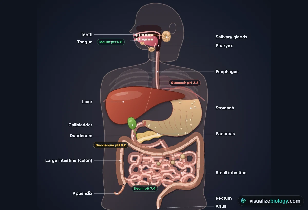
Parts of the digestive system and their functions
| Organ | What it does | Key facts |
|---|---|---|
| Mouth, teeth, tongue | Chews food (mechanical digestion), mixes it with saliva and forms a bolus | Salivary amylase: starch → maltose; pH about 7 |
| Salivary glands | Make saliva: water, mucus, amylase, lysozyme | 1–1.5 L a day |
| Esophagus | Carries the bolus to the stomach by peristalsis | 25 cm, about 8 s; no digestion |
| Stomach | Stores and churns food; acid kills microbes; pepsin starts protein digestion | pH 1.5–3.5; 2–4 h; makes chyme |
| Liver | Makes bile; processes absorbed nutrients; stores glycogen; makes urea | Largest internal organ, about 1.5 kg |
| Gallbladder | Stores and concentrates bile, releases it when fat arrives | Bile emulsifies fat; no enzymes |
| Pancreas | Makes amylase, trypsin, lipase and bicarbonate; insulin and glucagon | 1–1.5 L of juice a day, pH about 8 |
| Small intestine | Finishes digestion and absorbs nutrients through villi | Duodenum, jejunum, ileum; 6–7 m; about 30 m² of surface |
| Large intestine | Absorbs water and salts; gut bacteria ferment fibre | Cecum, appendix, colon; 1.5 m; 12–48 h |
| Rectum and anus | Store faeces and egest them | Two sphincters: one involuntary, one voluntary |
How digestion works, step by step
- Ingestion: food is taken into the mouth, chewed and mixed with saliva; amylase starts digesting starch.
- Swallowing: the epiglottis covers the windpipe and peristalsis, waves of muscle contraction, pushes the bolus down the esophagus.
- The stomach churns the food with hydrochloric acid and pepsin for 2–4 hours, turning it into chyme, and lets it out a little at a time.
- Digestion in the small intestine: bile emulsifies fat; pancreatic amylase, trypsin and lipase, and enzymes on the villi, finish the job.
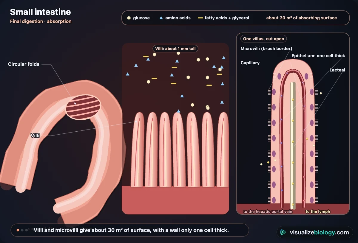
- Absorption: glucose and amino acids enter the blood capillaries of the villi; fatty acids and glycerol enter the lacteals.
- Assimilation: the liver and body cells use the absorbed molecules, for respiration, growth and repair.
- Egestion: the large intestine absorbs water, and the undigested remains leave through the anus as faeces.
Mechanical vs chemical digestion
Mechanical digestion breaks food into smaller pieces without changing its molecules: chewing, the churning of the stomach and the emulsifying action of bile. It gives enzymes a much larger surface area to work on. Chemical digestion is done by enzymes that break the bonds inside large molecules, making new, smaller molecules. Try it in the simulator: eat bread "gulped" and then "chewed well" and compare how fast starch is digested.
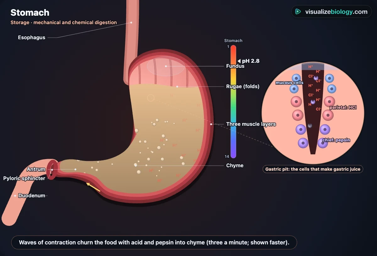
Digestive enzymes
| Enzyme | Made in | Works in | Breaks down → into | Best pH |
|---|---|---|---|---|
| Amylase | Salivary glands, pancreas | Mouth, small intestine | Starch → maltose | about 7 |
| Maltase | Small intestine lining | Small intestine | Maltose → glucose | about 6.5 |
| Pepsin (a protease) | Stomach | Stomach | Protein → peptides | about 2 |
| Trypsin (a protease) | Pancreas | Small intestine | Protein → peptides | about 8 |
| Peptidases | Pancreas, small intestine | Small intestine | Peptides → amino acids | about 7.5 |
| Lipase | Pancreas | Small intestine | Fats → fatty acids + glycerol | about 8 |
| Lactase, sucrase | Small intestine lining | Small intestine | Lactose, sucrose → simple sugars | about 6.5 |
Every enzyme works fastest at its optimum pH and at about body temperature; away from it, or above about 45 °C, the shape of its active site changes (it is denatured) and it stops working. That is why the pancreas sends bicarbonate to neutralize the acid chyme, and why the experiments here change pH and temperature.
How the small intestine absorbs food: villi
The lining of the small intestine is folded, the folds are covered in finger-like villi about 1 mm tall, and every cell on a villus has microvilli. Together they give a surface of about 30 m². Each villus has a wall only one cell thick, a dense network of blood capillaries that carry glucose and amino acids away to the liver in the hepatic portal vein, keeping a steep concentration gradient, and a lacteal that takes up fatty acids and glycerol into the lymph. The cells have many mitochondria to provide energy for active transport.
Follow the food: an interactive digestion animation
Choose a meal and press Play, and the camera rides with it through the body: chewed between the teeth for about half a minute, pushed down the 25 cm esophagus by peristalsis in about 8 seconds, churned in the stomach for 2–4 hours, squeezed through the pyloric sphincter a little at a time, carried along about 6–7 m of small intestine in roughly 3–4 hours, and dried out in the colon over a day or more. It is not a video: every mouthful is digested where it is, by enzymes that follow the local pH and temperature, so a fatty burger leaves the stomach later than toast, and a 75 g glucose drink raises blood glucose within minutes. Switch to Whole system to see the full canal at once, turn the 3D model to look from any side, or click an organ to see it alone, bigger and working.
A free digestion virtual lab
The Experiment mode is a digestion virtual lab that runs in the browser with no login: the classic practicals students do with test tubes and a water bath, with results computed from the same enzyme model as the simulation. Each one has a method checklist, a timer, a conclusion question and a PDF lab report.
- Amylase and pH: starch and amylase in buffers from pH 4 to 9, iodine on a spotting tile every 10 seconds, rate = 1000 ÷ time.
- Enzymes and temperature: the same reaction in water baths from 10 to 60 °C, to find the optimum and see denaturation.
- Lipase and bile: milk, lipase and phenolphthalein with and without bile, to show emulsification.
- Visking tubing model gut: why starch has to be digested before it can pass through a membrane.
- Food tests: iodine, Benedict's, biuret and the emulsion test on an unknown food.
- Surface area of the small intestine: folds × villi × microvilli, and what celiac disease does to it.
Experiments you can do
| Practical | What it shows | Course links |
|---|---|---|
| Parts of the digestive system | The organs and the order food passes through them | NGSS MS-LS1-3; IGCSE 0610 topic 7 |
| Food tests | Iodine for starch, Benedict's for reducing sugars, biuret for protein, the emulsion test for lipids | AQA GCSE required practical 3 |
| pH and amylase | Rate of starch digestion against pH, with a spotting tile and iodine | AQA GCSE required practical 4 |
| Temperature and amylase | The optimum temperature and denaturation | AP Biology 3.3; IGCSE; HS-LS1 |
| Lipase and bile | Bile emulsifies fat and speeds up lipase (phenolphthalein) | GCSE, IGCSE, A-level |
| Visking tubing model gut | Why starch must be digested before it can be absorbed | GCSE, IGCSE, NCERT |
| Surface area of the small intestine | Folds × villi × microvilli; celiac disease | A-level 3.3.3; IB |
Who uses this simulator?
Middle school, high school and early college students use it to see what a flat diagram cannot show: the bolus being squeezed down the esophagus, the stomach churning, bile breaking fat apart, villi taking up glucose and the colon drying out the waste. Teachers project it and ask "what will happen if…" before pressing Play (a fatty meal, no bile, lactose intolerance), and set the practicals when there is no time or equipment for them. It is an educational model built from published physiology, not medical advice; it needs no login and runs on school Chromebooks.
Frequently asked questions
What are the organs of the digestive system and their functions?
The alimentary canal is a tube about 9 m long. In the mouth, teeth break food up and saliva starts digesting starch. The esophagus carries the bolus to the stomach by peristalsis. The stomach churns food with hydrochloric acid and pepsin, which starts protein digestion. The small intestine (duodenum, jejunum and ileum, about 6–7 m) finishes digestion and absorbs nutrients through its villi. The large intestine absorbs water and holds gut bacteria, and the rectum stores faeces until they leave through the anus. Three accessory organs help without food passing through them: the liver makes bile, the gallbladder stores it, and the pancreas makes digestive enzymes and bicarbonate (and the hormones insulin and glucagon).
How does the digestive system work step by step?
1. Ingestion: food is taken into the mouth, chewed (mechanical digestion) and mixed with saliva, whose amylase starts digesting starch. 2. Swallowing: the tongue pushes the bolus into the pharynx, the epiglottis covers the windpipe and peristalsis carries it down the esophagus. 3. The stomach churns the food with acid and pepsin for 2–4 hours, turning it into chyme. 4. In the duodenum, bile emulsifies fat and pancreatic juice adds amylase, trypsin, lipase and bicarbonate; enzymes on the villi finish the job. 5. Absorption: glucose and amino acids pass into the blood capillaries of the villi and fatty acids and glycerol into the lacteals. 6. Assimilation: the body's cells use the absorbed molecules. 7. The large intestine absorbs water, and 8. egestion removes the undigested remains as faeces, usually a day or two after the meal.
What do amylase, protease and lipase do?
They are digestive enzymes that break large insoluble food molecules into small soluble ones that can be absorbed. Amylase (from the salivary glands and pancreas) digests starch into maltose, which maltase on the villi splits into glucose. Proteases digest proteins into peptides and amino acids: pepsin works in the acid stomach (best at about pH 2) and trypsin in the small intestine (about pH 8). Lipase (from the pancreas) digests fats into fatty acids and glycerol, helped by bile, which emulsifies the fat into tiny droplets with a large surface area. Each enzyme has an optimum pH and temperature (about 37–40 °C in the body); too hot or the wrong pH changes the shape of its active site and denatures it.
Is there a free digestive system virtual lab?
Yes. This simulator includes a free digestion virtual lab that runs in the browser with no sign-up: the effect of pH and temperature on amylase, lipase with and without bile, a Visking tubing model gut, food tests and the surface area of the small intestine. Each practical has a method, a timer, a conclusion question and a PDF lab report, and the results come from a model of real enzyme behaviour.
Can I see the digestive system in 3D?
Yes. Switch the simulator to 3D to turn the whole digestive system and look at it from any side: the mouth with its teeth, tongue and salivary glands, the esophagus, stomach, liver, gallbladder, pancreas, small and large intestines, rectum and anus. Click an organ and the others fade while the camera flies to it, and press Play to watch a meal move through the 3D organs.
Explore related simulators
See where the glucose you absorb ends up in the Virtual Living Cell, follow the blood that carries it from the intestine in the Heart Simulator, and watch water cross membranes, as it does in the colon, in the Osmosis Lab. Meet the gut's bacteria in the Virtual Bacteria Simulator, see how a single cell digests its food in a food vacuole in the Virtual Amoeba Simulator, and look at real tissue sections down the Virtual Microscope. Where does our food's energy come from in the first place? Follow it from the sun in Build the Food Chain. Breathe in the oxygen that releases that energy, and breathe out its carbon dioxide, in the Respiratory System Simulator. Follow what happens to the glucose from a meal once it is absorbed, as insulin and glucagon take over, in the Endocrine System Simulator. See the same organs in an amphibian, with a much shorter carnivore's gut, in the Virtual Frog Dissection.
Sources: OpenStax Biology 2e, chapter 34; OpenStax Anatomy and Physiology 2e, chapter 23; Urry et al., Campbell Biology 12th edition, chapter 41; Hall, Guyton and Hall Textbook of Medical Physiology 14th edition, chapters 63–66; Elashoff et al. (1982) Gastroenterology 83:1306; Helander and Fändriks (2014) Scand. J. Gastroenterol. 49:681; Lewis and Heaton (1997) Scand. J. Gastroenterol. 32:920. The model is an educational simplification of these sources; time is speeded up and the close-ups are not to scale.
Updated