
Virtual Frog Dissection: Frog Morphology and Anatomy, Step by Step
Pin a frog to the tray, cut it open with the scissors and lift out its organs one layer at a time, naming each one as you go. Then watch its heart, lungs, gut, kidneys and nerves at work in a see-through frog.
What you're looking at
Lab notebook
Every step of your dissection, each cut's accuracy, each organ lifted out and every measurement is written here. Export a CSV, or turn the session into a PDF lab report.
| # | Specimen | What | Result | Value |
|---|
Concept library
Click "New problem" to begin.
Click "Start quiz" for 5 questions on the frog's morphology, mouth, organs, digestion, breathing and blood, excretion and reproduction, nerves and calculations.
1 Overview
Simulate puts a frog on a dissecting tray, on the left, in four views: External (its morphology, from above and below), Mouth (held open), Dissection (pin it, cut it open, lift the organs out) and Systems (see-through, with one organ system working). On the right, How it works plays animated close-ups (heart, brain, urogenital system, breathing, digestion, life cycle) and Graph plots heart rate, oxygen uptake and gut length. Experiment has five practicals that end in a PDF lab report, and Quiz has a practice quiz with worked solutions and a 5-question quiz.
2 External features and the mouth
- Tap any part to read what it is, its job and an exam fact in What you're looking at; hover (on a computer) for its name. ▶ plays a guided tour.
- ↻ Flip over turns the frog to see its belly. Tap the eye to see the nictitating membrane; tap a male's throat to make him call.
- In the Frog panel choose the species (leopard frog, Indian bullfrog, common frog, American bullfrog) and the sex.
- In Mouth, Lift tongue shows that the tongue is attached at the front.
3 Doing the dissection
- Pick 📍 Pins (the right instrument is picked for you at each step) and tap the four yellow rings at the hands and feet.
- With ✂️ Scissors, press on the yellow dot and drag along the yellow line. A cut that strays or stops short is not accepted; each cut's accuracy goes in the notebook.
- Tap the belly with 🤏 Forceps to fold back the skin.
- Cut the muscle beside the dark blue ventral abdominal vein, then up the pectoral girdle.
- Tap organs with the forceps to lift them into the dish, top layer first, or use the Organs panel. 🔍 Hand lens magnifies; 📏 Ruler measures.
▶ Watch it done performs and explains the whole dissection; Cut for me does one step.
4 Systems and close-ups
Tap any organ (in Systems or in the dissection) to open its Organ page on the right: a labelled close-up, every part an exam asks for, what it does, why it matters to the frog, NCERT/NEET points and a comparison with humans. The chips under it step through the organs of the system. In Systems, pick an organ system in the Systems panel (or ⏭ Next system). With Show it working on, food, air, blood, urine and nerve impulses move. The temperature slider and Resting · Active · Hibernating change the heart rate and breathing in the close-ups and graphs.
5 Experiment mode and the lab report
Five practicals: external features and sexing, inside the mouth, the full dissection (graded), gut length (adult vs tadpole) and heart rate vs temperature. Each shows its aim, what you need and a real-lab safety note; the method ticks off as you work, a conclusion question checks your result, and Create lab report builds a PDF. The controls for each practical are in the Lab panel.
6 Units, keyboard and gestures
- SI | US switches between millimetres and degrees Celsius and inches and degrees Fahrenheit.
- Space plays or pauses, T does the view's next step, 1–4 choose the views, S opens Status, B counts a beat in the heart practical and Esc clears the selection.
- A plain scroll over the tray scrolls the page. Right-click the tray or the close-up to save it as an image.
Virtual frog dissection: the frog's morphology and anatomy, step by step
The frog has been the classic first dissection in biology for a century, because its body plan is a clear model of a vertebrate: a three-chambered heart, lungs and a skin that breathes, a digestive tract with a liver and pancreas, kidneys, gonads and a brain with cranial nerves. Today many schools offer an alternative to dissecting real animals, and NCERT's Class 11 chapter teaches the frog mostly from diagrams. This virtual frog dissection lets you do every step yourself on screen, with the organs drawn where they really sit, and then shows each organ system working.
- Specimen
- Northern leopard frog, Indian bullfrog (NCERT Rana tigrina), common frog or American bullfrog; male or female
- You do
- Pin, cut the skin and muscle, open the girdle, lift out organs with forceps, measure with a ruler
- Shows
- External features, the mouth, 40+ organs and structures, 7 organ systems at work
- Modes
- Simulate, Experiment (5 practicals with a PDF report) and Quiz
- Price
- Free, with no account or download; works on Chromebooks, tablets and phones
Frog dissection steps
- External features. Look at the frog dorsal side up, then ventral side up, and decide its sex.
- The mouth. Cut the angles of the jaws and find the teeth, nares, Eustachian tubes, glottis and gullet.
- Pin. Lay the frog on its back and pin it through the hands and feet.
- Skin incisions. Lift the skin with forceps; cut up the midline from the cloaca to the lower jaw, then across to each arm and leg; pin the flaps back.
- Muscle incisions. Cut the muscle wall the same way but just to one side of the midline, so you miss the ventral abdominal vein. Cut through the pectoral girdle to reach the heart.
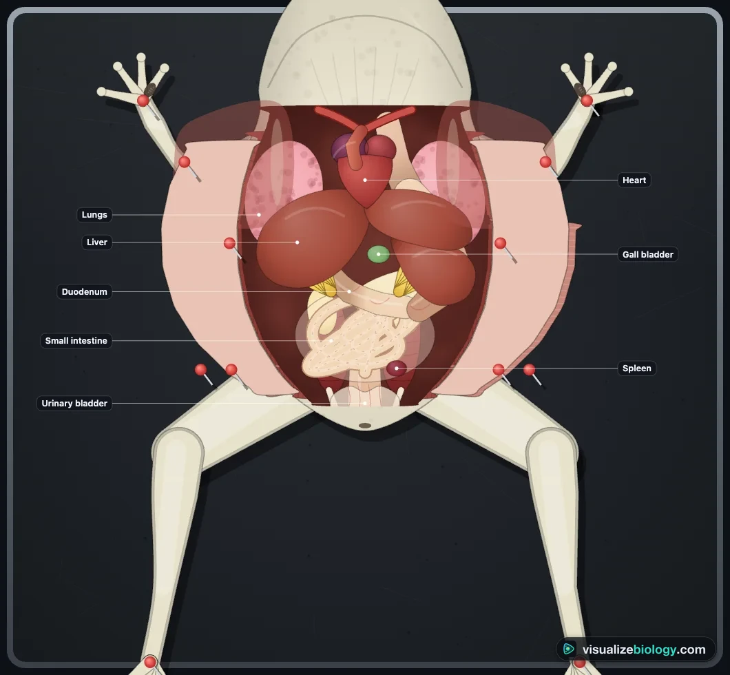
- Organs in place. Identify them, then lift out the fat bodies, liver, eggs (in a female) and the digestive tract to see the kidneys, gonads and the back wall.
Morphology of the frog: external features
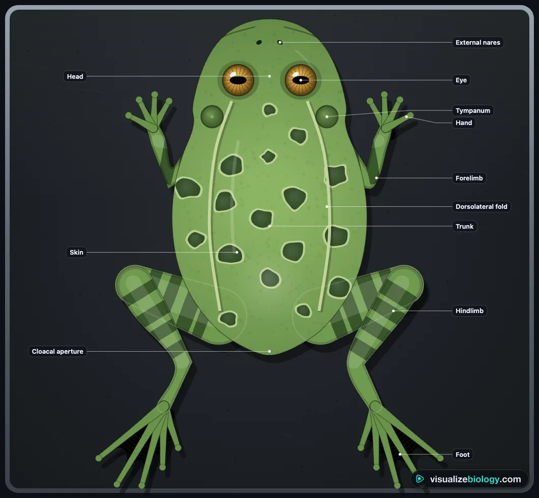
A frog's body has only a head and a trunk: no neck and no tail. Its skin is smooth and moist with mucus, olive green with dark spots above and pale below; a frog never drinks, it absorbs water through its skin. The bulging eyes have a see-through nictitating membrane; behind each eye is the tympanum, the eardrum, with no outer ear. Forelimbs have four digits; the long hindlimbs have five webbed toes. Males have vocal sacs and a nuptial (copulatory) pad on the first finger; females have neither.
Inside the frog's mouth
The maxillary teeth line the upper jaw and two patches of vomerine teeth sit on the roof; the lower jaw has none. The teeth only grip, because frogs swallow prey whole. The sticky tongue is attached at the front and forked at the back, so it flips out over the lower jaw. The internal nares lead to the nostrils, the Eustachian tubes to the ears, the glottis to the lungs and the gullet to the esophagus.
Internal anatomy of a frog: organs and their functions
| Organ | Where you find it | Function |
|---|---|---|
| Heart | Between the forelimbs, under the girdle | Two atria and one ventricle pump blood round a closed circulation |
| Lungs | Beside the heart, partly under the liver | Pulmonary respiration on land |
| Liver (3 lobes) and gall bladder | The largest organ, over the stomach | Makes bile, stored in the gall bladder; stores glycogen |
| Stomach | On the frog's left, J-shaped | HCl and gastric juice begin digestion; food leaves as chyme |
| Small intestine (duodenum, ileum) | Coiled in the mesentery | Receives bile and pancreatic juice; absorbs food through villi |
| Pancreas and spleen | Between stomach and duodenum; in the mesentery | Pancreatic juice and insulin; stores blood and removes old red cells |
| Fat bodies | Yellow, finger-like, on the kidneys | Energy for hibernation and breeding |
| Kidneys and ureters | On the back wall beside the backbone | Make urine (urea: frogs are ureotelic) |
| Urinary bladder and cloaca | At the hind end | Store urine; one exit for faeces, urine and gametes |
| Testes or ovaries | On the kidneys or filling the cavity | Make sperm or eggs |
The frog's three-chambered heart
Oxygen-poor blood from the body collects in the sinus venosus and enters the right atrium; oxygen-rich blood from the lungs enters the left atrium. Both empty into the single ventricle, which pumps blood into the conus arteriosus on the ventral side and out through three pairs of arches: carotid, systemic and pulmocutaneous. Frogs also have a hepatic portal and a renal portal system. Because a frog is an ectotherm, its heart rate rises with temperature, which you can measure in the heart-rate practical.
Male vs female frog
| Male | Female | |
|---|---|---|
| Outside | Vocal sacs; nuptial pad on the first finger | No vocal sacs or nuptial pads |
| Gonads | Yellowish testes held on the kidneys by the mesorchium | Large ovaries full of eggs, not joined to the kidneys |
| Ducts | 10–12 vasa efferentia → Bidder's canal → urinogenital duct → cloaca | Coiled oviducts open into the cloaca separately from the ureters |
Frog anatomy for NCERT Class 11 and NEET
NCERT's chapter on structural organisation in animals now studies only the frog, Rana tigrina. Its favourite facts are all in this simulator: the frog breathes through its skin in water and during hibernation and aestivation; it has 10 pairs of cranial nerves; its brain has a forebrain, midbrain and hindbrain; it is ureotelic; a female lays 2500–3000 eggs at a time and fertilisation is external, followed by a tadpole and metamorphosis.
Practicals and course links
| Practical | What you find out | Course links |
|---|---|---|
| External features and sexing | The frog's morphology, male or female | NCERT 11 ch. 7; high school biology |
| Inside the mouth | The structures of the buccal cavity | High school frog dissection labs |
| The full dissection | How the organs are arranged, in layers | NGSS HS-LS1-2; AP Biology |
| Gut length | Why a carnivore's gut is short and a tadpole's long | NCERT 11; AP Biology |
| Heart rate and temperature | Q10 in an ectotherm | AP Biology; A-level |
Who uses this virtual frog dissection?
High school students in the US doing the frog dissection lab (or its alternative), NCERT Class 11 students and NEET candidates learning the morphology and anatomy of the frog, AP and college biology students revising vertebrate organ systems, and teachers who want a free, no-login dissection on a classroom screen or students' Chromebooks.
Frequently asked questions
How do you tell a male frog from a female frog?
From outside, a male frog has vocal sacs at the throat, which blow up when he calls, and a dark, rough nuptial (copulatory) pad on the first finger of each hand; a female has neither. In some species, such as the American bullfrog, the male's tympanum is also wider than his eye. Inside, a male has two yellowish testes on the front of the kidneys, and his ureters carry both urine and sperm (urinogenital ducts); a female has large ovaries full of black-and-white eggs and long, coiled oviducts that open into the cloaca separately from the ureters.
What is the function of the nictitating membrane in frogs?
The nictitating membrane is a thin, see-through third eyelid that moves up over the frog's eye from below. It protects and moistens the eye under water and on land while the frog can still see through it.
How many cranial nerves does a frog have?
A frog has 10 pairs of cranial nerves, which leave the brain to serve the head (smell, sight, hearing, taste and the jaw muscles). Reptiles, birds and mammals have 12 pairs. The frog's brain has a forebrain (olfactory lobes, cerebral hemispheres and diencephalon), a midbrain (optic lobes) and a hindbrain (cerebellum and medulla oblongata).
What are the steps of a frog dissection?
Study the external features first and open the mouth to see its structures. Then lay the frog ventral side up and pin it through the hands and feet. Lift the skin with forceps and cut along the midline from the cloaca to the lower jaw, then across to each arm and leg, and pin the flaps back. Cut the muscle wall the same way but just to one side of the midline to miss the ventral abdominal vein, then cut through the pectoral girdle. Identify the organs in place, then lift out the fat bodies, liver, any eggs and the digestive tract to see the kidneys and gonads.
Explore related simulators
Compare the frog's three-chambered heart with our four chambers in the Heart & Cardiac Cycle Simulator, and its organ systems with ours in the Human Digestive System Simulator and the Kidney Simulator. Follow a frog's eggs from fertilisation in the Meiosis Simulator, see where it sits in a pond food web in Build the Food Chain, compare its metamorphosis from tadpole to frog with a caterpillar's in the Butterfly Life Cycle Simulator, dissect an insect for comparison in the Virtual Cockroach Dissection, and look at frog blood cells and tissues down the Virtual Microscope.
Sources: NCERT Biology Class 11 (2022 rationalised edition), chapter 7.2, Frogs; OpenStax Biology 2e, chapter 29.3 (Amphibians); Hutchison, Whitford and Kohl, Physiological Zoology 41 (1968), and Feder and Burggren, Biological Reviews 60 (1985), for skin and lung gas exchange; Gosner (1960) for development. Heart rates, gut lengths and development times are typical values for an educational model; real frogs vary with species, size, season and temperature.
Updated