
Kidney and Urinary System Simulator
An interactive, animated model of working kidneys that filter your blood and make urine: pressure pushes fluid out in the glomerulus, the nephron takes back what the body needs, ADH decides how much water to keep, and urine travels down the ureters to the bladder. Drink, sweat or eat salt and see what changes.
Experiment
08:00making 1.3 mL of urine a minute
Live readings
Filtration (GFR)—
Urine made—
Urine concentration—
Blood concentration—
ADH—
Blood pressure—
Blood glucose—
Bladder—
Eat, drink, move
The person
Blood going to the kidneys
Lab
View
The body's chemistry runs on the clock you choose (one second can be a minute or ten minutes). The flow you see in the tubes is slowed down and the ureter's waves sped up so both can be followed; the nephron and the glomerulus are enlarged thousands of times.
Clock
What the kidneys are doing
Lab notebook
Urine samples, drinks, measurements and test results are written here. Export a CSV, or turn the session into a PDF lab report.
| # | Time | Measured | Value | Notes |
|---|
Concept library
Click "New question" to begin.
Click "Start quiz" for 5 questions on the urinary system, the nephron, urine formation, ADH and the path of urine.
User Guide: Kidney Simulator
1 Overview
Simulate shows working kidneys in 3D (desktop) or 2D (phones), driven by one model of the body's water, salt, glucose and hormones. The body's clock runs as soon as the page opens: urine is made, flows down the ureters and fills the bladder. Choose a view on the left of the picture and a close-up, the urine sample or a graph on the right. Experiment has seven practicals that end in a PDF lab report, and Quiz has a practice quiz with worked answers and a 5-question quiz.
2 The five views
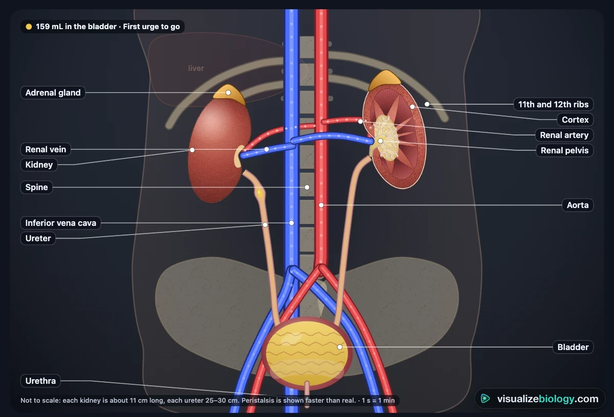
- Urinary system: both kidneys with the aorta, vena cava and renal vessels, the ureters (watch the waves of peristalsis carry urine down), the bladder filling, and the urethra (switch Female or Male in the Person panel).
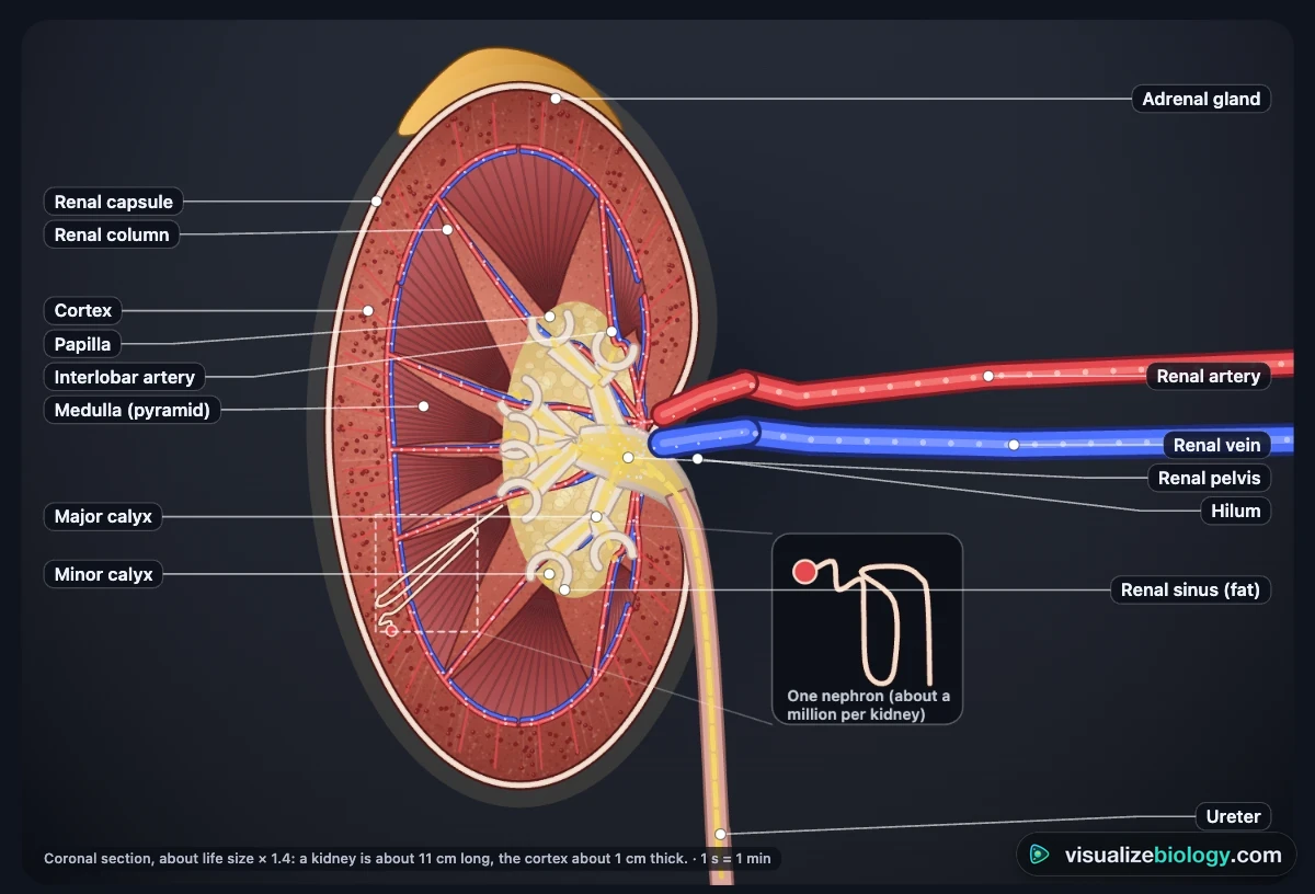
- Inside a kidney: a kidney cut lengthways: capsule, cortex, medulla and pyramids, papillae, calyces, renal pelvis and the blood vessels, with urine dripping into the calyces.
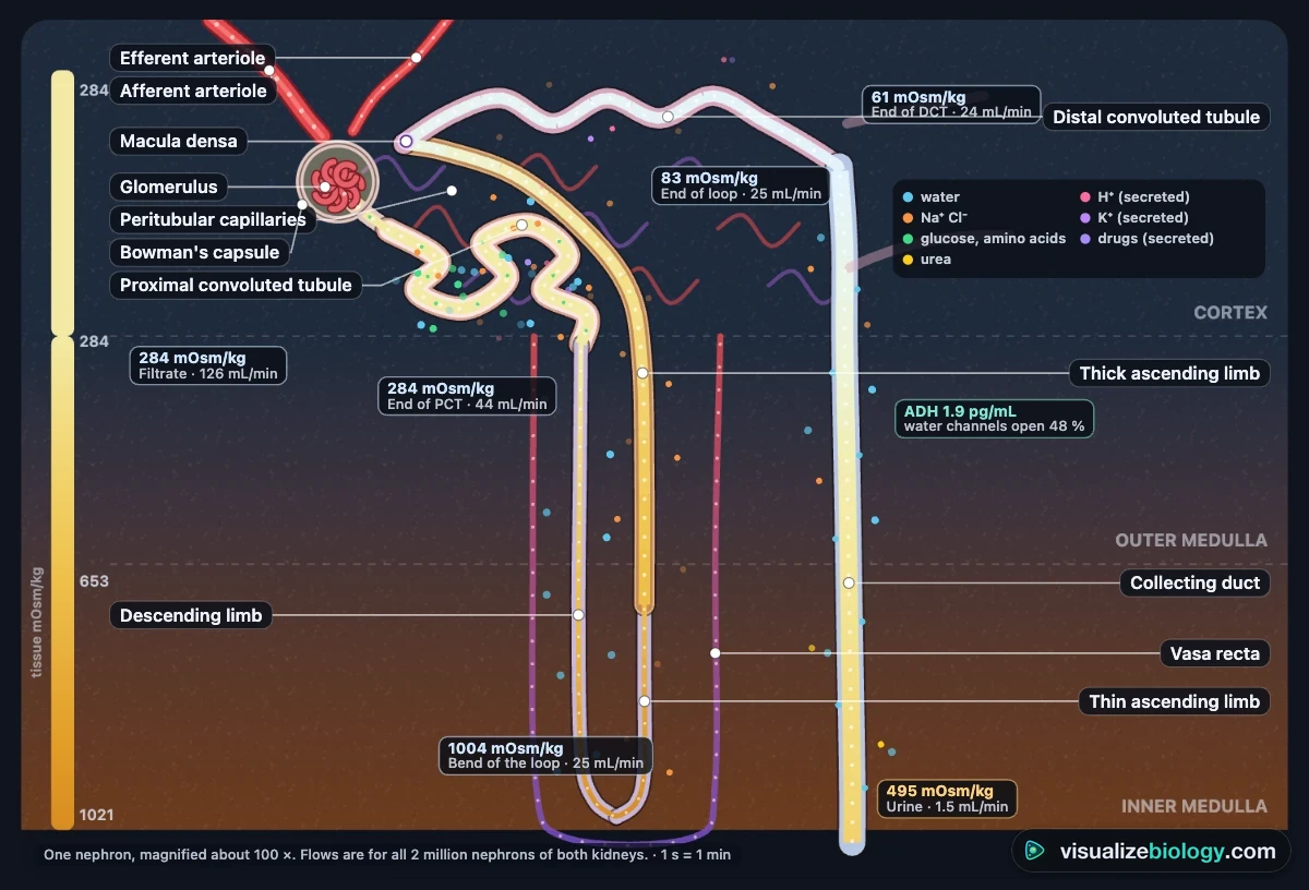
- Nephron: one nephron from the glomerulus to the collecting duct, in the cortex and the medulla, whose saltiness is shaded. Arrows show what leaves and enters each part; the tags give the concentration and the flow.
- Glomerulus: ultrafiltration close up, with the pressures that push fluid out and the cells and proteins that stay behind.
- Bladder: the bladder wall, sphincters and urethra, the stretch receptors and the nerves of the micturition reflex.
3 Eat, drink and move
In the Do panel give the person a glass of water, 500 mL or 1 L, a salty snack, a coffee, a beer, a sugary drink, an hour of exercise in the heat or a diuretic tablet, then watch the urine flow and concentration on the Graph (Time) and the color of the urine sample. Drinks when thirsty lets the person top up when the blood gets concentrated; turn it off to see dehydration. Go to the toilet empties the bladder; leave it too long and the reflex wins.
4 The person's condition and blood
The Person panel (or the Kidneys menu in the command bar) switches between healthy kidneys and diabetes mellitus, diabetes insipidus, nephrogenic diabetes insipidus, a kidney stone, a urinary tract infection, glomerulonephritis, chronic kidney disease, blood loss, high blood pressure and an enlarged prostate. The Blood panel can hold the blood pressure or the blood glucose at a value you choose, as in an experiment. These are educational models of typical cases, not medical advice.
5 The story (▶) and the clock
Press the round ▶ under the kidneys to play the story: about two minutes in which the camera follows one drop of plasma from the renal artery through the glomerulus and every part of the nephron, into the calyces and down the ureter in a wave of peristalsis, into the bladder and out through the urethra. It moves between the 3D model and the textbook section, a banner says what is happening at each place, and the close-up beside it shows the same place: the filter, the tubule cells, the urine. Use ‹ and › to skip, ❚❚ to pause and ✕ to stop. The body's clock waits while the story plays.
In Experiment mode the same button runs the body's clock. Run in the bar below starts or pauses the clock in any mode, and the speed pills set how fast it runs: Real time, 1 min/s or 10 min/s (two hours after a drink pass in 12 seconds).
6 Experiment mode and the lab report
Seven practicals: parts of the urinary system, a kidney dissection, water and salt and urine output, a urine test (dipstick) on a patient, the glucose threshold, blood pressure and filtration, and kidney dialysis. Each shows its aim, what you need and a safety note; the method ticks off as you work, a conclusion question checks your result, and Create lab report builds a PDF with your results, pictures and calculations. The tools of each practical are in the Lab panel.
7 Quiz and units
The practice quiz covers the parts of the urinary system, the nephron, ultrafiltration, reabsorption and secretion, ADH and water balance, the path of urine, kidney disease and treatment, and calculations (GFR, clearance, percentages), each with a worked answer. Take the quiz gives five mixed questions and a star rating. SI | US switches pressures between kPa and mmHg, glucose between mmol/L and mg/dL, creatinine between µmol/L and mg/dL and lengths between centimeters and inches. Volumes are in milliliters in both, as in every clinic.
8 Keyboard and gestures
- Space or F plays or pauses the story (in Experiment mode, Space runs the clock), ← → skip through it, 1–5 choose the view, W gives a glass of water, T goes to the toilet, L labels, C, U, G the close-up, urine and graph, S Status, Esc stops the story.
- In 3D, drag to turn the model and right-drag to move it; a plain scroll over it scrolls the page, so hold Ctrl or ⌘ (or click the model first) to zoom. On a phone one finger scrolls the page and two fingers turn the model.
- Right-click the picture or the close-up to save it as an image or copy the readings.
Kidney function, animated: structure, urine formation and the path of urine
Your two kidneys filter all the plasma in your blood about 60 times a day. They take out urea and other wastes, and keep the amount of water, salt and acid in the blood just right, so they are organs of excretion and of homeostasis at once. This simulator lets you watch them do it: see the pressure that forces fluid out in a glomerulus, follow the filtrate through a nephron, change how much the person drinks and see ADH respond, and follow the urine down the ureters to the bladder.
- Subject
- The human kidneys and urinary system: kidney, nephron, ureters, bladder, urethra
- It shows
- Ultrafiltration, selective reabsorption, secretion, the loop of Henle, ADH and aldosterone, peristalsis, urination
- Views
- Urinary system, inside a kidney, nephron, glomerulus, bladder; cell close-ups, urine test, graphs
- Modes
- Simulate (▶ plays a story that follows one drop), Experiment (7 practicals) and Quiz
- Price
- Free, no account or download; works on Chromebooks, tablets and phones
A kidney virtual lab, not a video
A video of urine formation plays the same way every time. Here the nephron answers what you do. Give the person a liter of water, a salty snack, a coffee, a beer or a diuretic, and watch ADH, the collecting duct and the urine change minute by minute. Raise or lower blood pressure to see filtration hold steady, then fail. Turn up blood glucose until sugar spills into the urine. The loop of Henle animation builds the salty medulla as a countercurrent multiplier, and pressing ▶ plays a story in which the camera follows one drop of plasma from the renal artery to the urethra. The Experiment mode adds a virtual kidney dissection, a GFR and blood-pressure practical, a urine test on a mystery patient and dialysis. It does the job of a renal physiology virtual lab, free, with no license or sign-up.
The parts of the urinary system and their functions
| Part | What it does |
|---|---|
| Kidneys | Filter the blood, remove urea and excess water and salt, and make urine; about 11 cm long, beside the spine under the lowest ribs |
| Renal artery and vein | Bring blood to each kidney (about 1.1 L a minute, a fifth of the heart's output) and take the cleaned blood away |
| Ureters | Two muscular tubes, 25–30 cm long, that carry urine from the kidneys to the bladder by peristalsis |
| Bladder | A stretchy muscular bag that stores urine; you feel the urge at about 150–200 mL and it holds about 500 mL |
| Sphincters | Rings of muscle that keep the bladder shut: the internal one works by reflex, the external one you control |
| Urethra | Carries urine out of the body: about 4 cm long in women and 20 cm in men |
The structure of the kidney
Cut a kidney lengthways and you see three regions. The outer cortex, about 1 cm thick and grainy, holds the glomeruli and the coiled tubules. The inner medulla is made of cone-shaped renal pyramids, striped by the loops of Henle and collecting ducts, with their tips (papillae) pointing inwards. Urine drips from each papilla into a calyx, and the calyces join into the renal pelvis, a funnel that becomes the ureter at the hilum, where the renal artery enters and the renal vein leaves. A tough capsule and a cushion of fat protect the whole kidney.
The nephron: structure and function
Each kidney has about a million nephrons, the tiny tubes that make urine. Every part of a nephron has its own job:
| Part | Where | What it does |
|---|---|---|
| Glomerulus and Bowman's capsule | Cortex | Ultrafiltration: blood pressure squeezes water and small molecules out of the blood |
| Proximal convoluted tubule | Cortex | Reabsorbs all the glucose and amino acids and about 65 % of the water and salt |
| Loop of Henle | Dips into the medulla | Makes the medulla salty, so water can be drawn out of the collecting duct |
| Distal convoluted tubule | Cortex | Fine-tunes salt (aldosterone), secretes potassium and hydrogen ions |
| Collecting duct | Cortex to papilla | Reabsorbs water under the control of ADH, then carries urine to the pelvis |
How urine is formed: ultrafiltration, selective reabsorption and secretion
- Ultrafiltration. The afferent arteriole is wider than the efferent one, so blood pressure in the glomerulus is high (about 55 mmHg). Against it push the fluid already in Bowman's capsule (15 mmHg) and the pull of the plasma proteins (about 30 mmHg), leaving a net 10 mmHg that forces water, glucose, amino acids, salts and urea through the filter. Blood cells and proteins are too big. Together the kidneys filter about 125 mL a minute, 180 liters a day.
- Selective reabsorption. The body needs most of that back. The proximal tubule, lined with microvilli and packed with mitochondria, reabsorbs all the glucose (by co-transport with sodium) and amino acids, and about two thirds of the salt and water. If blood glucose is above about 10 mmol/L (180 mg/dL), the carriers are full and glucose appears in the urine, a sign of diabetes.
- The loop of Henle. The ascending limb pumps salt out but is waterproof, so the medulla becomes salty, up to 1200 mOsm/kg at the tips of the pyramids: a countercurrent multiplier. Water then leaves the descending limb and the collecting duct by osmosis.
- Secretion. The tubules add hydrogen ions, potassium, ammonium and some drugs (such as penicillin) from the blood, which is how the kidneys control blood pH.
About 99 % of the filtered water is reabsorbed. What remains, about 1.5 liters a day, is urine: water with urea, salts, creatinine and other wastes.
ADH and osmoregulation
The water potential of the blood is kept steady by negative feedback. Osmoreceptors in the hypothalamus sense how concentrated the blood is, and the posterior pituitary gland releases more or less ADH (antidiuretic hormone). ADH makes the collecting duct cells put aquaporin water channels into their membranes, so more water is reabsorbed. A second hormone, aldosterone from the adrenal glands, controls sodium in the distal tubule and collecting duct when blood volume or pressure falls.
| Dehydrated (sweating, salty food) | Too much water (a big drink) | |
|---|---|---|
| Blood | Too concentrated | Too dilute |
| ADH from the pituitary | More | Less |
| Collecting ducts | More permeable: more water reabsorbed | Less permeable: less water reabsorbed |
| Urine | Small volume, concentrated, dark | Large volume, dilute, pale |
The path of urine
- Nephron and collecting duct → the papilla at the tip of a pyramid.
- Minor calyx → major calyx → renal pelvis.
- Out of the kidney at the hilum into the ureter; waves of peristalsis push it down a few times a minute.
- Into the bladder through slanted openings that stop it flowing back; the bladder stretches as it fills.
- Stretch receptors signal the spinal cord and brain. When you choose to go, the detrusor muscle contracts, the internal and external sphincters relax, and urine leaves through the urethra.
Kidney disease, dialysis and transplants
A urine test (dipstick) tells a doctor a lot: glucose suggests diabetes, protein and blood suggest damaged glomeruli, white cells and nitrite an infection. Kidney stones are crystals that can block a ureter and cause severe pain. In chronic kidney disease, usually caused by diabetes or high blood pressure, nephrons are lost for good; when fewer than about one in seven are left, urea and salts build up and the person needs dialysis or a transplant. In hemodialysis, blood flows past dialysis fluid on the other side of a partially permeable membrane, in the opposite direction (countercurrent), so urea keeps diffusing out while glucose and useful salts, already in the fluid at normal blood levels, stay. A transplanted kidney works all the time but needs drugs that stop the immune system rejecting it.
Experiments you can do
| Practical | What it shows | Course links |
|---|---|---|
| Parts of the urinary system | Kidney, ureter, bladder, urethra, renal vessels | NGSS MS-LS1-3; GCSE; IGCSE excretion |
| Kidney dissection | Cortex, medulla, pyramids, pelvis, ureter and vessels | GCSE, IGCSE, high school biology |
| Water, salt and urine output | ADH and negative feedback over time | GCSE 4.5.3.3; HS-LS1-3; AP Bio 2.8 |
| Urine test: diagnose the patient | Dipstick results and what they mean | GCSE, OCR A-level urine tests |
| The glucose threshold | Transport maximum, glucose in the urine in diabetes | A-level, IB, AP |
| Blood pressure and filtration | Ultrafiltration and autoregulation of GFR | A-level, IB, intro college |
| Kidney dialysis | Diffusion across a membrane; countercurrent flow | GCSE dialysis; A-level |
Who uses this simulator?
High school and early college students use it to see what a diagram cannot show: filtrate moving and shrinking along the nephron, the medulla getting saltier, ADH opening water channels, a wave of peristalsis carrying urine down a ureter. Teachers project it and ask "what will happen if…" before pressing a button (a liter of water, a salty snack, a marathon on a hot day, a kidney stone), and set the practicals when there is no kidney to dissect or no urine to test. It is an educational model built from published physiology, not medical advice; it needs no login and runs on school Chromebooks.
Frequently asked questions
How do the kidneys make urine?
Each kidney has about a million nephrons, and each makes urine in three steps. Ultrafiltration: blood pressure in the glomerulus, a knot of leaky capillaries, squeezes water, glucose, amino acids, salts and urea out into Bowman's capsule (about 180 liters a day), while blood cells and proteins are too big to pass. Selective reabsorption: the proximal convoluted tubule takes back all the glucose and amino acids and most of the water and salt, the loop of Henle makes the medulla salty, and the collecting duct takes back as much water as ADH allows. Secretion: the tubule adds hydrogen ions, potassium and some drugs. What is left, about 1.5 liters a day, is urine: water, urea, salts and other wastes.
What is the path of urine through the urinary system?
Urine forms in the nephrons and flows through the collecting ducts to the renal papillae at the tips of the pyramids. It drips into the minor calyces, then the major calyces and the renal pelvis, and leaves the kidney at the hilum through the ureter. Waves of muscle contraction (peristalsis) push it down each ureter, about 25 to 30 cm, to the urinary bladder, which stores it. When the bladder is full, stretch receptors send signals to the spinal cord and brain; during urination the detrusor muscle contracts, the internal and external urethral sphincters relax, and urine leaves the body through the urethra. In short: kidney, ureter, bladder, urethra.
What does ADH do in the kidney?
ADH (antidiuretic hormone, vasopressin) controls how much water the kidneys keep. When the blood becomes too concentrated, for example after sweating or eating salty food, osmoreceptors in the hypothalamus detect it and the pituitary gland releases more ADH. ADH makes the cells of the collecting ducts insert aquaporin water channels, so more water leaves the filtrate by osmosis into the salty medulla and back into the blood: a small volume of concentrated, dark urine is made. When you drink a lot, less ADH is released, the collecting ducts stay waterproof and a large volume of dilute, pale urine is made. This negative feedback keeps the water potential of the blood steady (osmoregulation).
Explore related simulators
See the heart that pumps a fifth of its output through the kidneys in the Heart Simulator, and how water moves by osmosis across a membrane in the Osmosis Lab, the same process that draws water out of the collecting ducts. Measure the blood pressure that drives filtration in the glomerulus with a cuff and a mercury column in the Manual Blood Pressure Apparatus Simulator. Look inside the cells that line the tubules in the Virtual Living Cell, and see how a single-celled organism gets rid of water with a contractile vacuole in the Virtual Amoeba Simulator. Plants move water too: compare with the Xylem and Phloem Simulator. The lungs share the kidneys' job of keeping the blood's pH steady: watch breathing blow off carbon dioxide in the Respiratory System Simulator. See ADH and aldosterone alongside the body's other hormones, and how negative feedback keeps them in check, in the Endocrine System Simulator.
Sources: OpenStax Biology 2e, chapter 41 (Osmotic Regulation and Excretion); OpenStax Anatomy and Physiology 2e, chapter 25 (The Urinary System); Urry et al., Campbell Biology 12th edition, chapter 44; Boron and Boulpaep, Medical Physiology 3rd edition, chapters 33–38; Hall, Guyton and Hall Textbook of Medical Physiology 14th edition, chapters 26–30; KDIGO (2012) CKD guideline; Michaels (1966) on dialyzer performance. The model is an educational simplification of these sources, not medical advice; flows are slowed down and the nephron is enlarged so they can be seen.
Updated