
Respiratory System Simulator: How the Lungs Breathe and Exchange Gas
Watch the diaphragm and ribs move, follow the air from the nose to the alveoli and the oxygen into the blood. Click any part to see it close up, then exercise, hold your breath, climb Everest or run a spirometer.
Experiment
Breathing12 breaths/min
Live readings
Breathing rate—
Tidal volume—
Ventilation—
SpO₂—
Arterial PO₂—
Arterial PCO₂—
Blood pH—
Heart rate—
Activity
Air and altitude
The body
Look at a part
Lab
View
Breathing runs in real time (12 breaths a minute at rest); ¼× and ½× slow it down, 5× speeds up exercise and recovery. The air is drawn far slower than it really moves, and the close-ups are not to scale.
Speed
What the body is doing
Lab notebook
Breathing readings, forced breaths, breath holds and practical measurements are written here. Export a CSV, or turn the session into a PDF lab report.
| # | Body | Measured | Value | Notes |
|---|
Concept library
Click "New question" to begin.
Click "Start quiz" for 5 questions on the airways, breathing, gas exchange, gas transport and lung health.
User Guide: Respiratory System Simulator
1 Overview
Simulate shows the human respiratory system breathing in real time, in 2D (or 3D with the 2D | 3D switch). The diaphragm and ribs move, the lungs fill and the air flows in and out of the airways; the pressure gauge shows the pressure in the lungs and the pleura against the outside air, and the magnifier shows the alveoli loading oxygen. Click any part to see it close up and working, while the right-hand card explains what it does. Experiment has seven practicals that end in a PDF lab report, and Quiz has a practice quiz with worked answers and a 5-question quiz.
2 Looking at one part
- Click (or tap) a part on the body, a label, or a button in the Parts panel. The view zooms in and the close-up plays: air warming in the nasal cavity, the epiglottis and vocal cords, the cilia carrying mucus up the trachea, the bronchial tree (and a bronchiole in asthma), the alveoli and one alveolar wall, the lungs' lobes and pleura, the diaphragm and ribs with Boyle's law, hemoglobin and the dissociation curve, and the brainstem's control of breathing.
- The Info card shows what the part is, how it works step by step, its key facts and what can go wrong, with a "Right now" line from the live model. Click a labelled structure inside a close-up to name it.
- ← Whole body (or Esc) zooms back out. In 3D the other parts fade so the chosen one is alone; drag to turn it.
3 Breathing: activity and actions
- The Breathe panel sets the activity (rest, walk, jog, run hard): the muscles use more oxygen and make more carbon dioxide, and breathing follows. Use the 5× speed to reach the new steady state, and to watch the recovery afterwards.
- Deep breath fills the lungs to total lung capacity; Breathe out fully empties them to the residual volume; Blow out hard records a forced expiration (FEV1, FVC and peak flow) on the flow–volume graph.
- Hold breath takes a big breath and holds it until you press again or the rising carbon dioxide forces a breath (the breaking point). Hyperventilate breathes fast and deep: blood CO₂ falls and the blood becomes alkaline. Try holding your breath straight afterwards, and watch the oxygen.
- ▶ / ❚❚ (Space) pauses and resumes breathing; ¼× and ½× slow it down.
4 Air, altitude and conditions
The Air panel takes the body to Denver, a mountain hut at 3,500 m or the summit of Everest, adds a 40 % oxygen mask, or acclimatizes the body for three weeks. The Body panel (or the Body menu in the command bar) gives it asthma (with a reliever inhaler), COPD, pulmonary fibrosis, pneumonia, a collapsed lung, a smoker's airways, anemia or carbon monoxide poisoning. Each changes the model and the close-ups. These are educational models of typical cases, not medical advice.
5 Spirometer, gases and graphs
- Spirometer draws the lung volume over the last 30 seconds on the lung volumes and capacities (TV, IRV, ERV, RV; IC, FRC, VC, TLC).
- Gases shows the partial pressures of oxygen and carbon dioxide from the air to the alveoli, the arteries, the tissues and the veins, and the make-up of breathed-in, breathed-out and alveolar air.
- Graph: the breathing cycle (volume, alveolar and pleural pressure, flow), the O₂–hemoglobin curve with the blood's points and the Bohr shift, blood gases and exercise over 5 minutes, the capnogram and the flow–volume loop.
- The Status button (key S) shows the live readings: breathing rate, tidal volume, ventilation, oxygen saturation, arterial PO₂ and PCO₂, pH and heart rate.
6 Follow a breath
Press 🧭 on the transport bar to follow an oxygen molecule in through the nose, down the airways, into the blood and to a working muscle, and the carbon dioxide back out. Each step opens a close-up. Use ‹ and › to step, ❚❚ to pause and Done to finish.
7 Experiment mode and the lab report
Seven practicals: the parts of the respiratory system, the bell jar model (drag the rubber sheet), measuring lung volumes with a spirometer (click two points on the drum to measure between them), exercise and breathing, carbon dioxide in exhaled air with bromothymol blue, spirometry for four patients (read FEV1 and FVC off the curve), and the oxygen–hemoglobin dissociation curve with a tonometer. Each shows its aim, what you need and a safety note; the method ticks off as you work, a conclusion question checks your result, and Create lab report builds a PDF. The apparatus is in the Lab panel.
8 Quiz and units
The practice quiz covers the parts, the mechanism of breathing, lung volumes and calculations, gas exchange, gas transport, the control of breathing and lung health, each with a worked answer. Take the quiz gives five mixed questions and a star rating. SI | US switches pressures between kPa and mmHg (the US textbook convention), heights between cm and inches and altitude between meters and feet. Volumes stay in mL and L in both, as on every spirometer.
9 Keyboard and gestures
- Space pauses or resumes breathing, D takes a deep breath, H holds the breath, 1–9 open the parts from the nose down, Esc goes back to the whole body, L labels, I see inside, F follow a breath, O, P, X, G the info card, spirometer, gases and graph, S Status.
- In 3D, drag to turn the body and right-drag to move it; a plain scroll over it scrolls the page, so hold Ctrl or ⌘ (or click it first) to zoom. On a phone one finger scrolls the page and two fingers turn the body.
- Right-click the body or the graph to save it as a picture or copy the readings.
The human respiratory system: how breathing and gas exchange work
Every cell in the body needs oxygen for aerobic respiration and makes carbon dioxide that must be removed. The respiratory system moves air in and out of the lungs and brings it so close to the blood that the two gases can swap places by diffusion. This respiratory system simulator lets you watch it happen: the diaphragm and ribs move, air flows from the nose to the alveoli, oxygen loads onto hemoglobin, and one physiological model keeps the blood gases steady as you exercise, hold your breath or climb a mountain.
- Subject
- The human respiratory system: airways, lungs, breathing muscles and gas transport
- It shows
- Breathing in real time, airflow, alveolar and pleural pressure, gas exchange, hemoglobin, control of breathing
- Views
- The whole system in 2D or 3D; animated close-ups of every part; spirometer, gases and graphs
- Modes
- Simulate (with Follow a breath), Experiment (7 practicals) and Quiz
- Price
- Free, no account or download; works on Chromebooks, tablets and phones
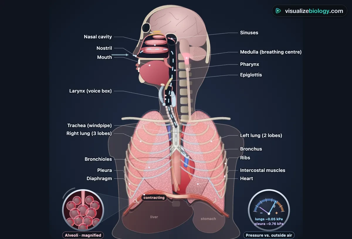
Parts of the respiratory system and the path of air
| Part | What it does | Key facts |
|---|---|---|
| Nose and nasal cavity | Warms, moistens and filters the air; smell | Three conchae; sticky mucus and cilia |
| Pharynx | The throat: shared by air and food | Naso-, oro- and laryngopharynx |
| Larynx | The voice box; the epiglottis closes it when you swallow | Vocal cords; thyroid cartilage (Adam's apple) |
| Trachea | The windpipe to the chest | About 12 cm; 16–20 C-shaped cartilage rings |
| Bronchi and bronchioles | Branch about 23 times to carry air through the lungs | Bronchioles have smooth muscle, no cartilage |
| Alveoli | Gas exchange with the blood | About 480 million; 70 m²; barrier 0.5 µm |
| Lungs and pleura | Hold the airways and alveoli; the pleura lets them slide and stay stretched | Right lung 3 lobes, left lung 2 |
| Diaphragm and intercostal muscles | Change the volume of the chest for breathing | The diaphragm does about 70 % of a quiet breath |
Inhalation vs exhalation: the mechanism of breathing
| Breathing in (inhalation) | Breathing out (exhalation, at rest) | |
|---|---|---|
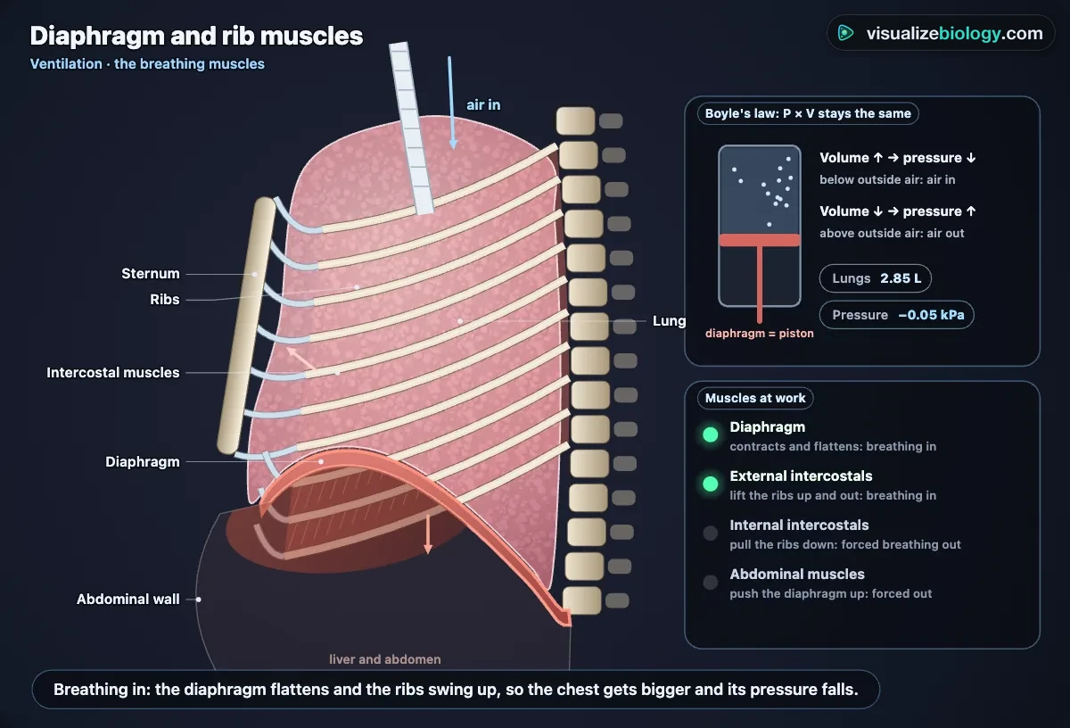
| Diaphragm | Contracts and flattens | Relaxes and domes up |
| External intercostals | Contract: ribs move up and out | Relax: ribs move down and in |
| Volume of the chest | Increases | Decreases |
| Pressure in the lungs | Falls below atmospheric (about −0.1 kPa) | Rises above atmospheric (about +0.1 kPa) |
| Air flows | In | Out |
The link between volume and pressure is Boyle's law: at a fixed temperature, pressure × volume stays the same. The lungs follow the chest wall because they are wrapped in the pleura, two thin membranes with a film of fluid between them; the pressure in that pleural space is below atmospheric (about −0.5 kPa). Forced breathing out also uses the internal intercostals and the abdominal muscles. In the simulator, watch the pressure gauge while the diaphragm moves.
The bell jar (balloon) lung model
In the classic model, a bell jar is the chest, two balloons on a Y-tube are the lungs, trachea and bronchi, and a rubber sheet stretched across the bottom is the diaphragm. Pulling the sheet down makes the jar bigger, its pressure falls and the balloons inflate; letting it go pushes the air back out. A hole in the jar (like a pneumothorax) stops the balloons inflating at all. The model has limits: the jar cannot move like the rib cage, and balloons are not lungs. Experiment mode lets you pull the sheet and read the pressure on a water manometer.
Gas exchange in the alveoli
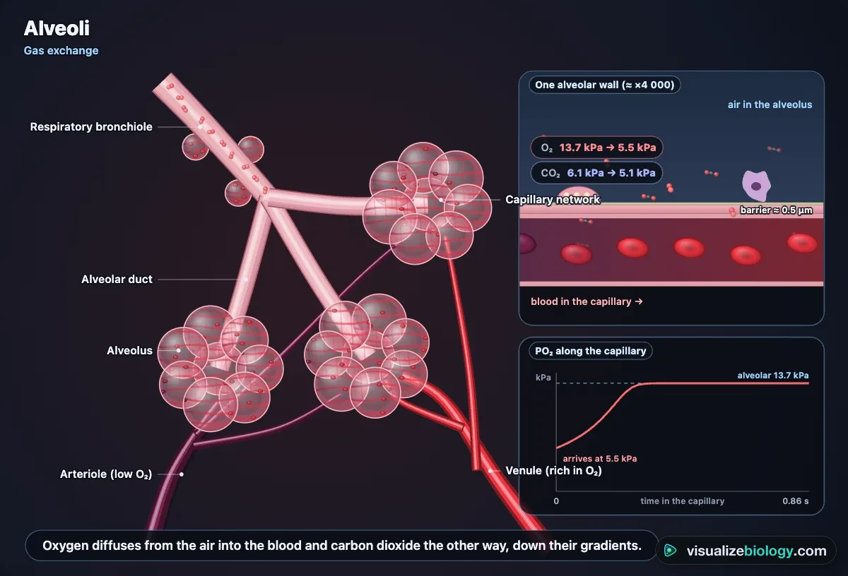
Oxygen diffuses from the air in the alveoli (PO₂ about 13.9 kPa) into the blood arriving from the pulmonary artery (about 5.3 kPa), and carbon dioxide diffuses the other way. Diffusion is fast because the alveoli have a huge surface (about 70 m²), a barrier only 0.5 µm thick (one flat type I cell, a basement membrane and one capillary cell), a dense capillary network, and breathing and blood flow keep the gradients steep. Type II cells make surfactant so small alveoli do not collapse. At rest blood reaches the alveolar PO₂ in about a quarter of a second, a third of its time in the capillary.
Lung volumes and capacities: tidal volume vs vital capacity
| Volume or capacity | Meaning | Adult man (woman) |
|---|---|---|
| Tidal volume (TV) | One quiet breath | 500 mL (500) |
| Inspiratory reserve volume (IRV) | Extra air you can breathe in | 3100 mL (1900) |
| Expiratory reserve volume (ERV) | Extra air you can breathe out | 1200 mL (700) |
| Residual volume (RV) | Always left in the lungs | 1200 mL (1100) |
| Vital capacity (VC = IRV + TV + ERV) | The biggest breath out after the biggest breath in | 4800 mL (3100) |
| Total lung capacity (TLC = VC + RV) | Air in fully inflated lungs | 6000 mL (4200) |
A spirometer measures these from a trace of volume against time; it cannot measure the residual volume. Minute ventilation is the breathing rate × tidal volume (about 12 × 0.5 L = 6 L/min at rest); about 150 mL of each breath only fills the airways (the dead space). In the forced test, FEV1 ÷ FVC below 70 % means obstructed airways, as in asthma and COPD.
The oxygen–hemoglobin dissociation curve and the Bohr effect
About 98 % of the oxygen in blood is carried by hemoglobin: four chains, each with an iron-containing heme that binds one O₂. Its saturation plotted against PO₂ is S-shaped, because each O₂ that binds makes the next bind more easily. In the lungs hemoglobin loads to about 98 %; in resting tissues it falls to about 75 %. In working muscle more CO₂, more acid and a higher temperature shift the curve to the right (the Bohr effect), so even more oxygen is released. Carbon dioxide returns mostly as hydrogen carbonate ions (about 70 %).
Control of breathing
The rhythm is set in the medulla of the brainstem and sent down the phrenic nerve to the diaphragm. Central chemoreceptors sense carbon dioxide (as acidity), the main drive: a small rise makes you breathe much more. The carotid and aortic bodies respond to low oxygen, which matters at altitude. This negative feedback keeps arterial CO₂ near 5.3 kPa, even during exercise, when ventilation can rise from 6 to over 100 L/min.
Breathing vs respiration
Breathing (ventilation) is moving air in and out of the lungs. Respiration has two meanings: gas exchange in the lungs and tissues (external and internal respiration), and cellular respiration, the reactions in mitochondria that use oxygen to release energy from glucose and make carbon dioxide and water. Breathing exists to supply cellular respiration.
Asthma, COPD, pneumothorax and other conditions
In asthma the smooth muscle round the bronchioles tightens and their lining swells, so breathing out is slow and wheezy; a reliever inhaler relaxes the muscle. In COPD and emphysema the walls between alveoli are destroyed, leaving less surface and trapped air. Fibrosis stiffens and thickens the lungs; pneumonia fills alveoli with fluid; in a pneumothorax air in the pleural space lets a lung collapse. Smoking paralyses the cilia, and carbon monoxide takes the place of oxygen on hemoglobin. The simulator shows each as an educational model of a typical case, not medical advice.
Experiments you can do
| Practical | What it shows | Course links |
|---|---|---|
| Parts of the respiratory system | The structures and the path of air, as in a lung dissection | NGSS MS-LS1-3, HS-LS1-2 |
| Bell jar (balloon) lung model | How the diaphragm changes volume and pressure; the model's limits | Middle school, GCSE, IGCSE |
| Lung volumes with a spirometer | TV, IRV, ERV, vital capacity, breathing rate, O₂ uptake | A&P lung volume lab, AP Biology |
| Exercise and breathing | Breathing rate, depth and ventilation with exercise and recovery | NGSS HS-LS1-3 (feedback) |
| CO₂ in exhaled air | Bromothymol blue turns yellow faster after exercise | Middle and high school |
| Spirometry and lung disease | FEV1, FVC and FEV1/FVC: obstructive vs restrictive; reversibility | A&P, intro college physiology |
| O₂–hemoglobin dissociation curve | The S-shaped curve, P50 and the Bohr shift | AP Biology, A-level, college |
Who uses this simulator?
High school and college students use it to see what a flat diagram cannot: the diaphragm and ribs moving together, air slowing as the airways branch, the pressures that make air flow, oxygen crossing into red blood cells and the brainstem raising breathing when CO₂ climbs. Anatomy and physiology classes use the spirometer, FEV1/FVC and dissociation-curve labs; teachers project it and ask "what will happen if…" before pressing Deep breath, Hold breath or Everest. It needs no login and runs on school Chromebooks.
Frequently asked questions
How does the diaphragm help you breathe?
The diaphragm is a dome of muscle under the lungs. When it contracts it flattens and moves down by about 1.5 cm in a quiet breath (up to about 10 cm in a deep one), while the external intercostal muscles lift the ribs up and out. The chest gets bigger, so the pressure inside the lungs falls just below the air outside (Boyle's law: pressure × volume stays constant) and air flows in. When the diaphragm relaxes it moves back up, the stretched lungs recoil, the pressure rises a little above atmospheric and air flows out. Quiet breathing out is passive; forced breathing out also uses the internal intercostals and abdominal muscles.
What is the difference between tidal volume and vital capacity?
Tidal volume is the air breathed in or out in one normal, quiet breath: about 500 mL in an adult at rest. Vital capacity is the largest volume you can breathe out after the deepest possible breath in: the tidal volume plus the inspiratory reserve volume (about 3100 mL) plus the expiratory reserve volume (about 1200 mL), so about 4.8 L in an adult man and 3.1 L in a woman. Even after breathing out fully, about 1.2 L, the residual volume, stays in the lungs, so the total lung capacity is about 6 L. Vital capacity rises with height and falls with age and in restrictive lung diseases such as fibrosis.
In the bell jar model, what do the balloons represent?
In the bell jar (balloon) model of the lungs, the two balloons represent the lungs, the Y-shaped glass tube represents the trachea and the two bronchi, the bell jar represents the rib cage (the chest cavity) and the rubber sheet at the bottom represents the diaphragm. Pulling the sheet down increases the volume of the jar, so the pressure of the air round the balloons falls and air from outside flows in and inflates them, just as in breathing in. The model has limits: the jar is rigid while the ribs move, balloons are not elastic tissue like lungs, and there is no pleural fluid.
Explore related simulators
Follow the oxygenated blood from the lungs to the body in the Heart Simulator, see oxygen used in mitochondria in the Virtual Living Cell, and watch diffusion and osmosis across a membrane in the Osmosis Lab. See how an insect breathes without lungs, through spiracles and tracheae, in the Virtual Cockroach Dissection. Measure how exercise raises blood pressure as well as breathing in the Manual Blood Pressure Apparatus Simulator. The medulla that sets your breathing is in the 3D Brain Model; the swallow that the epiglottis guards is in the Digestive System Simulator, and the kidneys that balance the blood's acid over days are in the Kidney Simulator. Look at real lung tissue sections down the Virtual Microscope.
Sources: OpenStax Anatomy and Physiology 2e, chapter 22; OpenStax Biology 2e, chapter 39; West and Luks, West's Respiratory Physiology 10th edition; Hall, Guyton and Hall Textbook of Medical Physiology 14th edition, chapters 38–43; Severinghaus (1979) J Appl Physiol 46:599; Hankinson, Odencrantz and Fedan (1999) Am J Respir Crit Care Med 159:179; West et al. (1983) J Appl Physiol 55:678. The model is an educational simplification of these sources; the air is drawn far slower than it moves and the close-ups are not to scale.
Updated