
Interactive 3D Brain Model: Lobes and Functions
Click any lobe and it splits out of the brain to show what it does. Then watch the brain at work, follow a signal from eye or hand to cortex, examine a stroke patient, and fire a neuron.
Experiment
Click a lobe to split it outor press ▶ to see the brain at work
Live readings
Selected—
Lobe—
Controls the body—
Blood supply—
Brodmann area—
Neurons—
Membrane potential—
Condition—
Split a lobe out
Or click a lobe on the brain. Click it again to choose one of its areas.
What is the brain doing?
The brain’s condition
A neuron
The Hodgkin–Huxley squid axon (1952): it lives in cold sea water and stops working above about 30 °C.
Lab
View
The brain is computed from one model: the same folds, lobes and areas in 2D and 3D. Activity is shown like an fMRI scan: the glow marks the areas working hardest, not every neuron that fires.
What the brain is doing
Lab notebook
Stimulation responses, bedside tests, reaction times, split-brain trials and touch thresholds are written here. Export a CSV, or turn the session into a PDF lab report.
| # | Brain | Measured | Value | Notes |
|---|
Concept library
Click "New question" to begin.
Click "Start quiz" for 5 questions on the lobes, the cortex, the deep brain, neurons and the methods used to study the brain.
User Guide: Interactive 3D Brain Model
1 Overview
Simulate shows a human brain in 3D (desktop) or 2D (phones), both computed from the same model with real gyri and sulci. Choose a view on the left of the brain, and read what the selected part does in the Functions tab on the right, beside a neuron, the homunculus and graphs. Experiment has seven practicals that end in a PDF lab report, and Quiz has a practice quiz with worked answers and a 5-question quiz.
2 Split a lobe out
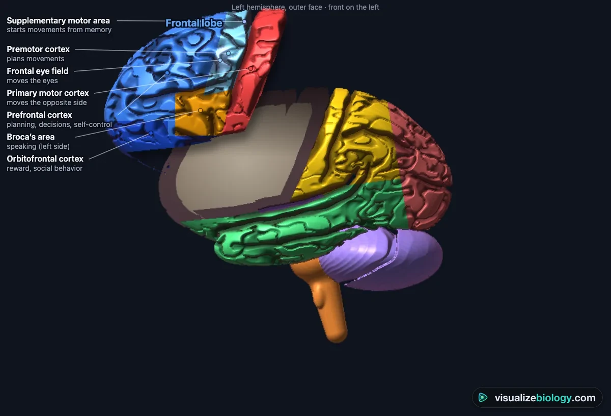
Click any lobe and it lifts out of the brain: its functional areas are colored and named with what each does, its cut face shows grey matter (the cortex) round white matter, and the Functions tab lists what the lobe does, its main areas, what happens when it is damaged and its blood supply. Click the lifted lobe again to choose one of its areas. Click outside the brain, press Esc or Put it back together to return it. ⤢ (or Pull every lobe apart) separates all the lobes at once; split the temporal or frontal lobe to see the insula hidden underneath. Left | Right chooses the hemisphere.
3 The five views
- Lobes: the outer surface in the lobes' colors (or real color), with the central sulcus and lateral fissure.
- Functional areas: motor, sensory, language, hearing, vision and association areas with their Brodmann numbers; the inner face is in the corner.
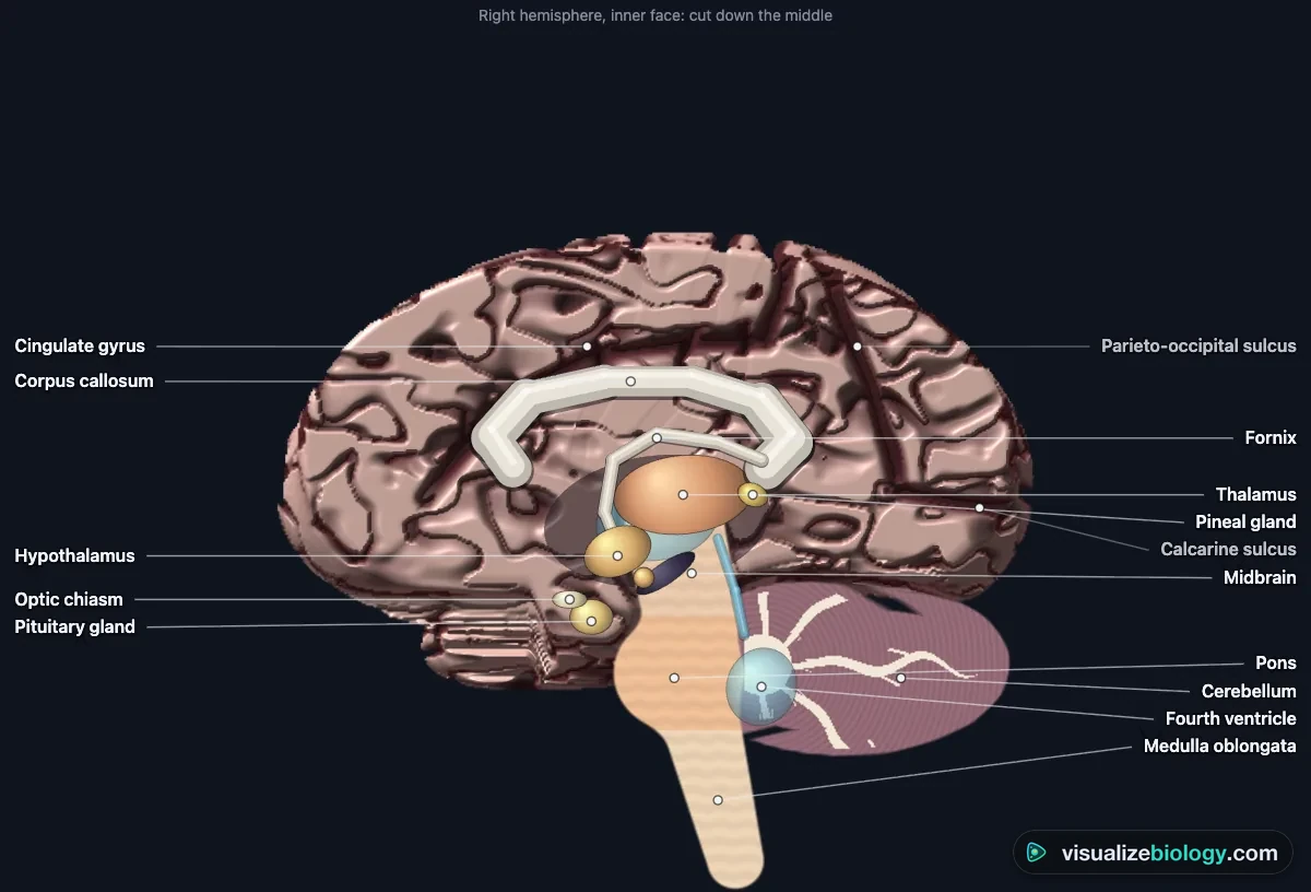
- Midline cut: the brain cut down the middle: corpus callosum, fornix, thalamus, hypothalamus, pituitary and pineal glands, the ventricles, midbrain, pons, medulla and the cerebellum.
- Deep inside: the limbic system (hippocampus, amygdala), the basal ganglia, the thalamus and the ventricles, seen through the brain.
- Blood supply: the territories of the anterior, middle and posterior cerebral arteries and the Circle of Willis.
4 Brain activity and Follow the signal
In the Tasks panel choose what the brain is doing (moving the right hand, feeling a touch, looking at a face, listening to music or speech, speaking, reading aloud, remembering, deciding, fear, balance, dreaming) and press ▶: the areas working hardest glow, as on an fMRI scan. 🧭 Follow the signal steps through the pathway for that task, from the senses or the decision to the muscles, with a spark that travels from one area to the next. Use ‹ and › to step, ❚❚ to pause and Done to finish.
5 Conditions
The Brain panel (or the Brain menu in the command bar) changes the brain: strokes of the left or right middle cerebral artery, the anterior and posterior cerebral arteries, Broca's and Wernicke's aphasia, Phineas Gage's frontal injury, cerebellar damage, Parkinson's and Alzheimer's diseases and the split brain. The damaged area darkens, and the panel lists the signs that follow from it. These are educational models of typical cases, not medical advice.
6 Neuron, homunculus and graphs
Neuron fires an action potential (the Hodgkin–Huxley model) and shows it travelling along the axon, jumping between the nodes of Ranvier when the axon is myelinated, with Na⁺ rushing in and K⁺ flowing out, and a synapse releasing its transmitter. Change the stimulus, the diameter, the myelin, the temperature and drugs (TTX blocks Na⁺ channels, TEA blocks K⁺ channels) in the Neuron panel. Homunculus draws each body part as big as its strip of motor or sensory cortex: tap one to light up its place on the brain. Graph shows the action potential, EEG rhythms, a night's sleep, conduction speed against diameter and reaction times.
7 Experiment mode and the lab report
Seven practicals: parts of the brain (as in a sheep brain dissection), brain mapping by stimulating the cortex (Penfield), examining a stroke patient to find the damage, the ruler-drop reaction time test, the split-brain experiment, two-point touch discrimination and reading an EEG. Each shows its aim, what you need and a safety note; the method ticks off as you work, a conclusion question checks your result, and Create lab report builds a PDF with your results, pictures and calculations. The controls for each practical are in the Lab panel.
8 Quiz and units
The practice quiz covers the lobes, the cortex and language, the deep brain and brainstem, neurons and impulse calculations, reaction time, EEG and sleep, and disorders, each with a worked answer. Take the quiz gives five mixed questions and a star rating. SI | US switches centimeters, meters per second, kilograms and °C to inches, feet per second, pounds and °F; brain sizes stay in millimeters and micrometers, as in every course.
9 Keyboard and gestures
- 1–5 choose the view, Space starts or stops the activity, F follows the signal, X pulls the lobes apart, Esc puts them back, H switches hemisphere, L labels, E fires the neuron, I, N, M, G the Functions, Neuron, Homunculus and Graph tabs, S Status.
- In 3D, drag to turn the brain and right-drag to move it; a plain scroll over it scrolls the page, so hold Ctrl or ⌘ (or click the brain first) to zoom. On a phone one finger scrolls the page and two fingers turn the brain.
- Right-click the brain or the close-up to save it as a picture or copy your results.
An interactive 3D brain model you can take apart
Most 3D brain models online let you turn the brain and read a label. This one comes apart. Click any lobe and it lifts out of the brain: its functional areas are colored and named, its cut face shows grey matter over white matter, and a Functions panel says what the lobe does, what happens when it is damaged and which artery feeds it. Click one of its areas for the detail, or click outside the brain to put the lobe back. Everything is free and runs in the browser, with no sign-up or download.
What you can do with the 3D brain
| Do this | What you see |
|---|---|
| Click a lobe | The frontal, parietal, temporal or occipital lobe, the insula, the cerebellum or the brainstem lifts out with its areas labeled |
| Drag to rotate, pinch or Ctrl + scroll to zoom | The brain from any side, with the central sulcus, lateral fissure and other real sulci |
| Open the midline view | The corpus callosum, thalamus, hypothalamus, ventricles, brainstem and the cerebellum’s arbor vitae |
| Switch the view | Lobes, functional areas with Brodmann numbers, deep structures (hippocampus, amygdala, basal ganglia) or the blood supply |
| Pick a task | The areas that light up when you move a hand, see a face, hear, speak, read aloud, remember or feel fear |
| Pick a condition | A stroke, aphasia, Parkinson’s disease or a split brain: the damaged area and the signs it causes |
The 3D model and the flat 2D diagrams are drawn from the same anatomical model, so a lobe you meet in 3D sits in the same place in every diagram. Phones open in 2D, where lobes split out the same way, and the 3D view is one tap away. Each lobe keeps one color in every view, so students building a brain model project from clay or foam can copy the lobes and the sulci between them straight off the screen.
Parts of the brain and their functions
The human brain weighs about 1.35 kg and holds some 86 billion neurons, each joined to thousands of others. It has three main parts. The cerebrum, the two wrinkled hemispheres, thinks, senses, moves the body, speaks and remembers. The cerebellum underneath it coordinates movement and balance. The brainstem joins the brain to the spinal cord and keeps us breathing.
- Subject
- The human brain: lobes, functional areas, deep structures, brainstem and cerebellum, neurons
- It shows
- Lobes splitting out with their functions, brain activity, signal pathways, strokes and other conditions, the action potential
- Views
- Lobes, functional areas, midline cut, deep inside, blood supply; neuron, homunculus, graphs
- Modes
- Simulate (with Follow the signal), Experiment (7 practicals) and Quiz
- Price
- Free, no account or download; works on Chromebooks, tablets and phones
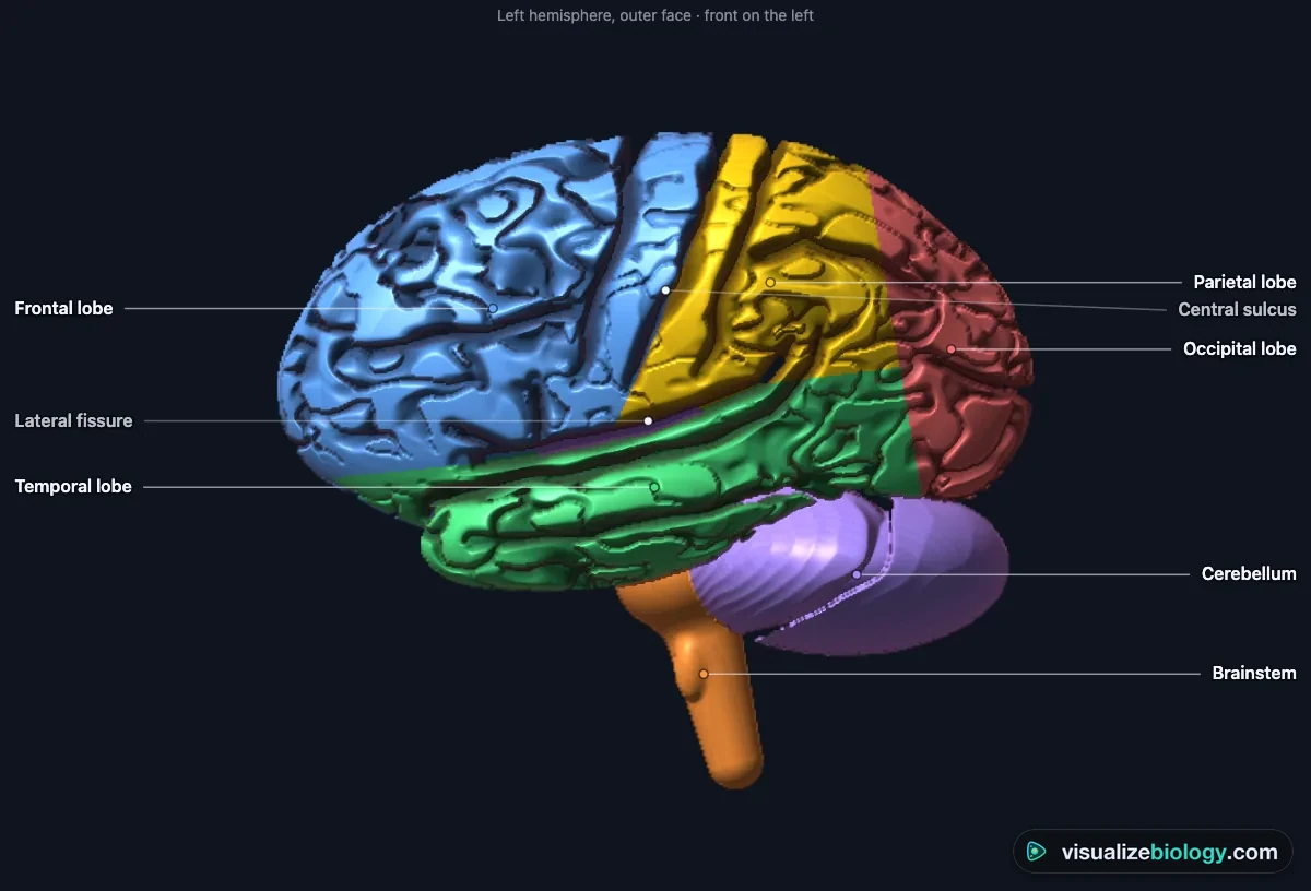
The lobes of the brain and their functions
| Lobe | Where | Main functions | If it is damaged |
|---|---|---|---|
| Frontal | In front of the central sulcus | Planning, decisions, personality, working memory; voluntary movement (primary motor cortex); speech (Broca’s area) | Weakness of the opposite side, personality change, Broca’s aphasia |
| Parietal | Behind the central sulcus | Touch, pain, temperature and body position (somatosensory cortex); spatial awareness; reading and arithmetic | Numbness of the opposite side, neglect of one side of space |
| Temporal | Below the lateral fissure | Hearing; understanding language (Wernicke’s area); memory (hippocampus); recognizing faces; emotion (amygdala) | Wernicke’s aphasia, memory loss, face blindness |
| Occipital | At the back | Vision: the primary visual cortex and areas for shape, color and motion | Loss of the opposite half of the visual field |
| Insula | Hidden in the lateral fissure | Taste, feelings from inside the body, disgust, cravings | Changed taste and body awareness |
| Cerebellum | Under the occipital lobes | Coordination, balance, posture, timing and motor learning | Clumsiness on the same side, unsteady walking, tremor |
| Brainstem | Midbrain, pons, medulla | Breathing, heart rate, blood pressure, swallowing, sleep and arousal; relays signals to the body | Life-threatening; locked-in syndrome, coma |
Inside the brain: from the corpus callosum to the brainstem
The cortex is a sheet of grey matter only 2–4 mm thick, folded into ridges (gyri) and grooves (sulci) so that about 2400 cm² fits in the skull; underneath is white matter, the insulated axons that connect areas. The corpus callosum, about 200 million axons, joins the two hemispheres. Below it the thalamus relays every sense except smell to the cortex, and the hypothalamus controls temperature, hunger, thirst and sleep, and the pituitary gland. The limbic system (hippocampus for memory, amygdala for fear) and the basal ganglia (choosing movements, with dopamine from the substantia nigra) lie deep inside. Four ventricles hold cerebrospinal fluid, which cushions the brain.
The motor cortex, sensory cortex and the homunculus
The primary motor cortex lies on the precentral gyrus, just in front of the central sulcus, and the primary somatosensory cortex on the postcentral gyrus just behind it. Each is a map of the opposite side of the body, with the toes at the top near the midline and the face and throat low down by the lateral fissure. Parts we control or feel finely, such as the hands, lips and tongue, get far more cortex than the back or legs: drawn to that scale, the body becomes the homunculus. Wilder Penfield found these maps in the 1940s by gently stimulating the cortex of awake patients during epilepsy surgery.
Left brain vs right brain, and the split brain
Each hemisphere moves and feels the opposite side of the body, because the pathways cross in the brainstem, and sees the opposite half of the visual field, because half the optic nerve fibers cross at the optic chiasm. In about 95 % of right-handers language is in the left hemisphere: Broca’s area in the frontal lobe for speaking and Wernicke’s area in the temporal lobe for understanding. The right hemisphere leads in attention to space, faces and the tune of speech. When the corpus callosum is cut to control epilepsy, a picture shown only to the left visual field reaches only the right hemisphere: the patient cannot name it, yet the left hand picks it out. The two hemispheres always work together, so "left-brained" and "right-brained" people are a myth.
How neurons send signals: the action potential
A neuron at rest is about −70 mV inside. When a stimulus pushes it past the threshold of about −55 mV, voltage-gated sodium channels open, Na⁺ rushes in and the inside swings to about +40 mV (depolarization); then potassium channels open, K⁺ flows out and the membrane repolarizes, briefly overshooting below rest. This action potential is all-or-none: a stronger stimulus makes impulses more frequent, not bigger. A myelin sheath makes the impulse jump between the nodes of Ranvier, up to about 120 m/s. At the axon’s end, the impulse releases a neurotransmitter across a synapse to the next cell.
Blood supply and stroke
The brain uses about 20 % of the body’s energy and receives about 750 mL of blood a minute. The anterior, middle and posterior cerebral arteries each supply their own territory, and they are joined underneath by the Circle of Willis. When one is blocked, the area it feeds stops working: a left middle cerebral artery stroke weakens the right face and arm and takes away speech, while a right one causes left-sided weakness and neglect. Spot a stroke with FAST: Face drooping, Arm weakness, Speech difficulty, Time to call emergency services.
Experiments you can do
| Practical | What it shows | Course links |
|---|---|---|
| Parts of the brain (dissection) | The lobes, cerebellum, brainstem and midline structures and their jobs | GCSE 4.5.2; IGCSE; HS biology |
| Brain mapping by stimulation | The motor and sensory strips and the homunculus | AP Psychology Unit 1; IB |
| Stroke patient | Locating damage from the signs: crossed control, language, neglect | Anatomy & physiology; college |
| Reaction time: ruler drop | Reaction time from t = √(2d ÷ g); practice, hand and cue | GCSE required practical; HS-LS1-3 |
| The split-brain experiment | Crossed visual pathways and left-hemisphere language | AP Psychology; IB |
| Two-point discrimination | Touch sensitivity and the sensory homunculus | GCSE; HS biology |
| Reading an EEG | Brain waves and the stages of sleep | AP Psychology; A-level; IB |
Who uses this 3D brain model?
High school and early college students use it to see what a flat diagram cannot: the lobes coming apart, the grey matter round the white, the hidden insula, the deep structures in place and an impulse jumping along a myelinated axon. Teachers project it to ask "what will happen if…" before revealing the answer (a stroke on the left, a cut corpus callosum, a blocked artery), and set the practicals when there is no sheep brain or EEG to hand. It is an educational model built from published anatomy and physiology, not medical advice; it needs no login and runs on school Chromebooks.
Frequently asked questions
What are the lobes of the brain and what do they do?
Each cerebral hemisphere has four lobes, plus the insula hidden inside the lateral fissure. The frontal lobe plans, decides and controls behavior and personality, holds the primary motor cortex that moves the opposite side of the body, and in the left hemisphere Broca's area for speaking. The parietal lobe, behind the central sulcus, feels touch, pain, temperature and body position and maps the space around us. The temporal lobe, below the lateral fissure, hears, understands language (Wernicke's area, usually on the left), forms new memories (hippocampus) and recognizes faces and objects. The occipital lobe at the back is for vision. The insula handles taste and feelings from inside the body. Below them, the cerebellum coordinates movement and balance and the brainstem controls breathing, heart rate and alertness.
What is the difference between the cerebrum, cerebellum and brainstem?
The cerebrum is the largest part: the two hemispheres with their folded cortex of grey matter over white matter, responsible for thinking, senses, voluntary movement, language and memory. The cerebellum is the smaller, finely folded 'little brain' under the back of the cerebrum; it coordinates movement, balance, posture and timing, and holds about 80 % of all the brain's neurons. The brainstem (midbrain, pons and medulla oblongata) joins the brain to the spinal cord; it controls automatic functions such as breathing, heart rate, blood pressure, swallowing and sleep, relays signals between brain and body, and carries most of the cranial nerves.
Why does the left side of the brain control the right side of the body?
Because the nerve pathways cross. Most motor fibers leaving the primary motor cortex cross to the other side in the medulla, at the decussation of the pyramids, before running down the spinal cord, and touch pathways also cross on their way up. So the left motor cortex moves the right side of the body and the left somatosensory cortex feels it. Vision crosses too: at the optic chiasm, fibers from the inner half of each retina cross, so the left hemisphere sees the right half of the visual field. The cerebellum is the exception: its pathways cross twice, so each side of the cerebellum coordinates the same side of the body.
Explore related simulators
Follow light to the visual cortex in the Human Eye Simulator and sound to the auditory cortex in the Human Ear Simulator, two sense organs that feed the brain, and see where touch, pain and temperature signals start in the Interactive Skin Diagram. See the pump that sends the brain its 750 mL of blood a minute in the Heart Simulator, look inside the animal cells that neurons are specialized from in the Virtual Living Cell, and examine real tissue down the Virtual Microscope. Take the skull that protects the brain apart, bone by bone, in the Interactive 3D Skeleton. See how the hypothalamus and pituitary run the body's hormones, from the thyroid loop to the stress response, in the Endocrine System Simulator.
Sources: OpenStax Anatomy and Physiology 2e, chapters 12–14; OpenStax Biology 2e, chapter 35; OpenStax Psychology 2e, chapter 3; Urry et al., Campbell Biology 12th edition, chapter 49; Hodgkin and Huxley (1952) J. Physiol. 117:500; Azevedo et al. (2009) J. Comp. Neurol. 513:532; Penfield and Rasmussen (1950) The Cerebral Cortex of Man; Weinstein (1968) two-point thresholds; Hursh (1939) conduction velocity. The model is an educational simplification of these sources; real brains vary from person to person.
Updated