
Interactive 3D Human Skeleton: All 206 Bones
Click any bone to see what it is, what it does and what it joins. Search for a bone and fly to it, explore each region, pull the skull apart and compare a male and a female skeleton.
Your exploration
Lab notebook
The bones you find, sort and count, your femur measurements, the traits of a mystery skeleton and the vinegar results are written here. Export a CSV, or turn the session into a PDF lab report.
| # | Skeleton | Measured | Value | Notes |
|---|
Concept library
Click "New question" to begin.
Click "Start quiz" for 5 questions: naming bones from pictures, where bones are, bone types, the functions and structure of bone, and male vs female skeletons.
1 Overview
Simulate shows the adult human skeleton in 3D (desktop) or as 2D textbook plates (phones), both made from the same anatomical model. Click any of the 206 bones and the Bone panel on the right shows what it is. Experiment has six practicals that end in a PDF lab report, and Quiz has a practice quiz with worked answers and a 5-question quiz.
2 Click a bone
Hover a bone to see its name, click it to select it. The Bone panel shows its name and everyday name, its type, its length (measured on the model), what it does, where it is, the bones it joins with the name and kind of each joint (click one to jump to that bone), its landmarks, a common injury, a fact and, where it matters, how it differs in men and women. ◎ Isolate (or double-click) shows the bone on its own, turning slowly, with its landmarks labelled. ⌂ or Esc goes back to the whole skeleton. For a bone with a partner on the other side, Left | Right switches to it.
3 Find a bone
Type in the search box above the Bone panel (or press /, or Find a bone in the command bar). Proper names (scaphoid), everyday names (collarbone, kneecap, shinbone, tailbone, funny bone, wisdom tooth), numbers (C1, T7, L5, rib 5) and small spelling slips all work. Pick a result and the view flies to the bone and highlights it. All bones lists every bone by region and group.
4 Regions and spotlights
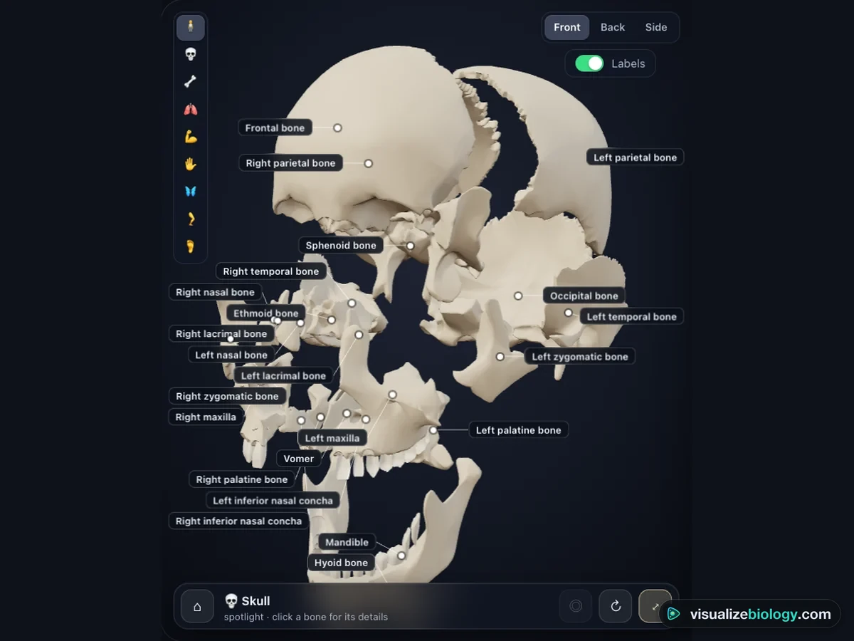
The rail on the left of the skeleton picks a region (skull, spine, chest, shoulder and arm, wrist and hand, pelvis, leg, ankle and foot): the camera frames it, the rest of the skeleton fades, and with Labels on every bone in it is named. The Spotlight row above the lab opens the major bones and sets on their own: the skull (press ⤢ to pull its 22 bones apart), the femur (with the inside of a long bone), the pelvis (with the male–female differences), the spine, the rib cage, the hand, the foot, and three vertebrae side by side.
5 Male and female
♂ Male | ♀ Female in the command bar changes the skeleton. The female skeleton is smaller and more slender, with a wider, lower pelvis, a wider subpubic angle, a shorter and wider sacrum, femurs that slope in more, smaller brow ridges and mastoids, and a more pointed chin. The Male vs female tab lists each trait with the values measured on the two models. Real people vary: any one trait overlaps between the sexes.
6 Colors and views
In the View panel, color the bones by Region or by Type (long, short, flat, irregular, sesamoid), or see them as an X-ray. Switch the costal cartilages and intervertebral discs, and the teeth, on and off. Front | Back | Side turns the skeleton (2D: chooses the plate). ↻ turns it slowly.
7 Experiment mode and the lab report
Six practicals: Name that bone (find bones on an unlabeled skeleton against the clock), Sort the bones by type, Count the bones of a hand and a foot, Height from a femur (measure it on an osteometric board and use the Trotter–Gleser formula), Male or female? (score the traits of a mystery skeleton) and Bones in vinegar (what acid and heat do to bone). Each shows its aim, what you need and a safety note; the method ticks off as you work, a conclusion question checks your result, and Create lab report builds a PDF. The controls for each practical are in the Lab panel.
8 Quiz and units
The practice quiz covers naming bones from pictures, where bones are and how many there are, bone types, the functions and structure of bone, joints, and male vs female skeletons with stature calculations, each with a worked answer. Take the quiz gives five mixed questions and a star rating. SI | US switches centimeters and grams to inches, feet and ounces; forensic measurements stay in millimeters, as in every osteology lab.
9 Keyboard and gestures
- / search, Esc or H whole skeleton, I isolate, R turn slowly, X pull the skull apart, M male or female, L labels, 1–9 regions, B, A, C the Bone, All bones and Male vs female tabs, S Status.
- In 3D, drag to turn the skeleton and right-drag to move it; a plain scroll over it scrolls the page, so hold Ctrl or ⌘ (or click the skeleton first) to zoom. On a phone one finger scrolls the page and two fingers turn and zoom.
- Right-click the skeleton to save it as a picture or copy your results.
An interactive 3D skeleton with all 206 bones
This is a free, interactive 3D model of the human skeleton that you can take bone by bone. Click any bone and a panel tells you what it is called (and what people call it: the collarbone, the kneecap, the tailbone), what type of bone it is, what it does, which bones it joins and through which joint, its landmarks and how it is most often injured. Search for a bone and the view flies to it. Every bone is a real anatomical mesh from BodyParts3D, not a drawing, so the shapes are those you would see on a real skeleton in a lab.
What you can do with the 3D skeleton
| Do this | What you see |
|---|---|
| Click a bone | Its name, type, length, job, joints, landmarks, injuries and a fact |
| Search | Any bone by name, everyday name or number (T7, rib 5), with the camera flying to it |
| Pick a region | The skull, spine, chest, arm, hand, pelvis, leg or foot, with every bone named |
| Open a spotlight | The skull pulled apart into 22 bones, the femur and the inside of a long bone, the pelvis, spine, rib cage, hand, foot and vertebrae |
| Switch to female | A wider pelvis and subpubic angle, a smoother skull, a smaller and more slender skeleton |
| Change the colors | Bones colored by region or by type, or an X-ray look |
The bones of the human body
An adult skeleton usually has 206 bones; a newborn has about 270 to 300, which fuse as the child grows. The skeleton supports the body, protects the organs, works with the muscles to move it, makes blood cells in the red marrow, and stores calcium and phosphate. It is about 15 % of body mass, and it is alive: bone is broken down and rebuilt so steadily that the whole skeleton is replaced about every ten years.
- Subject
- The adult human skeleton: all 206 bones, with cartilage, discs, teeth and ear ossicles
- It shows
- Every bone’s name, type, job, joints and landmarks; regions; spotlight views; male and female skeletons
- Views
- 3D (desktop), 2D textbook plates (phones); colors by region, by type, X-ray
- Modes
- Simulate, Experiment (6 practicals) and Quiz
- Price
- Free, no account or download; works on Chromebooks, tablets and phones
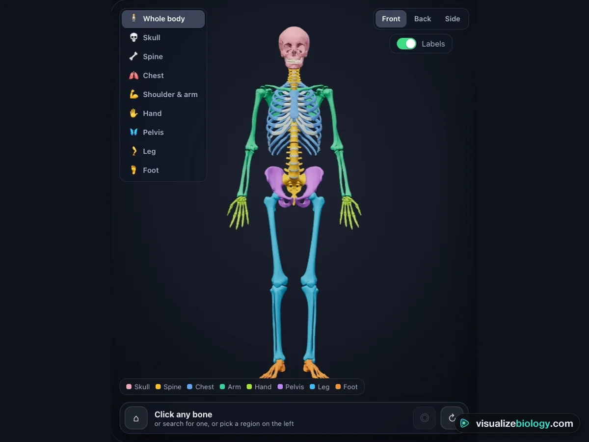
The axial and appendicular skeleton
| Part | Bones | Count |
|---|---|---|
| Skull | 8 cranial bones (frontal, 2 parietal, 2 temporal, occipital, sphenoid, ethmoid) and 14 facial bones (mandible, 2 maxillae, 2 zygomatic, 2 nasal, 2 lacrimal, 2 palatine, 2 inferior nasal conchae, vomer) | 22 |
| Ear ossicles and hyoid | Malleus, incus and stapes in each ear; the hyoid in the neck | 7 |
| Vertebral column | 7 cervical, 12 thoracic and 5 lumbar vertebrae, the sacrum and the coccyx | 26 |
| Rib cage | The sternum and 12 pairs of ribs (7 true, 3 false, 2 floating) | 25 |
| Shoulders and arms | Clavicle, scapula, humerus, radius and ulna on each side | 10 |
| Wrists and hands | 8 carpals, 5 metacarpals and 14 phalanges in each | 54 |
| Pelvis and legs | Hip bone, femur, patella, tibia and fibula on each side | 10 |
| Ankles and feet | 7 tarsals, 5 metatarsals and 14 phalanges in each | 52 |
The first four rows are the axial skeleton (80 bones), the body’s central axis that protects the brain, spinal cord, heart and lungs. The rest is the appendicular skeleton (126 bones): the limbs and the girdles that attach them, built for movement. More than half of all bones are in the hands and feet.
Types of bones
Bones are grouped by shape. Long bones (femur, humerus, phalanges) are levers, with a shaft of compact bone round a marrow cavity and wide ends of spongy bone. Short bones (the carpals and tarsals) are cube-shaped for stability. Flat bones (the skull roof, ribs, sternum, scapula) protect organs and give broad areas for muscles. Irregular bones (vertebrae, the hip bone, the sphenoid) fit no other shape, and sesamoid bones (the patella, the pisiform) grow inside tendons as pulleys.
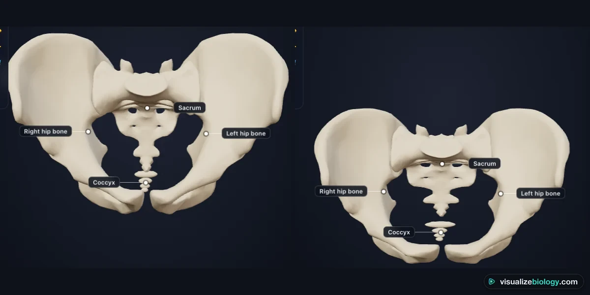
Male vs female skeleton
The pelvis tells the sexes apart best, because a woman’s pelvis is shaped for birth: it is wider and shallower, with a subpubic angle of about 80–90° (a U) instead of 50–60° (a V), a rounder inlet, a wider greater sciatic notch and a shorter, broader sacrum. On this model the subpubic angle measures 57° on the male pelvis and 85° on the female one. Male skulls usually have heavier brow ridges, bigger mastoid processes and a squarer jaw. Forensic anthropologists score several traits together and estimate height from the long bones: the femur is about a quarter of a person’s height.
Experiments you can do
| Practical | What it shows | Course links |
|---|---|---|
| Name that bone | The names and positions of the major bones | Middle school; GCSE; HS anatomy |
| Sort the bones by type | Long, short, flat, irregular and sesamoid bones | GCSE PE; HS anatomy |
| Count the bones | 27 bones in a hand and wrist, 26 in a foot and ankle | Middle school; IGCSE |
| Height from a femur | Stature from long-bone length (Trotter–Gleser) | Forensic science; HS biology |
| Male or female? | Sex traits of the pelvis and skull | Forensic science; college anatomy |
| Bones in vinegar | Mineral gives hardness, collagen gives flexibility | Middle school; GCSE; HS biology |
Who uses this 3D skeleton?
Middle school, high school and early college students use it to learn the bones and see where each one sits, instead of memorizing a flat diagram; teachers project it to quiz a class or to pull the skull apart in front of them, and use the practicals when there is no skeleton, femur or osteometric board in the room. Students of forensic science use the male and female skeletons and the height practical. It is an educational model built from published anatomy, not medical advice. It needs no login and runs on school Chromebooks.
Frequently asked questions
How many bones are in the human body?
An adult usually has 206 bones. A newborn baby has about 270 to 300, many of them still partly cartilage; as the child grows, several of them fuse together: the skull bones, the five vertebrae of the sacrum, the four of the coccyx and the three parts of each hip bone, for example. The number varies a little from person to person, because some people have an extra rib, extra small bones in the sutures of the skull or extra sesamoid bones in tendons. Of the 206, 80 make up the axial skeleton (skull, ear ossicles, hyoid, spine and rib cage) and 126 the appendicular skeleton (the shoulder and hip girdles and the limbs). More than half of them, 106, are in the hands and feet.
What is the difference between the axial and appendicular skeleton?
The axial skeleton is the central axis of the body: the skull (22 bones), the six ear ossicles, the hyoid bone, the vertebral column (26 bones) and the rib cage (the sternum and 24 ribs), 80 bones in all. It protects the brain, the spinal cord, the heart and the lungs and holds the body upright. The appendicular skeleton is made of the limbs and the girdles that attach them: the clavicles and scapulae (the shoulder or pectoral girdle), the bones of the arms and hands, the two hip bones (the pelvic girdle) and the bones of the legs and feet, 126 bones in all. It is built mainly for movement.
How can you tell a male skeleton from a female skeleton?
The pelvis is the most reliable guide, because a woman's pelvis is shaped for childbirth. A female pelvis is wider and shallower, with a wide subpubic angle (about 80 to 90 degrees, a U shape, against 50 to 60 degrees and a V in males), a round or oval inlet, a wide greater sciatic notch, ischial tuberosities further apart and a shorter, wider sacrum. The skull helps too: men usually have a larger brow ridge, bigger mastoid processes, a more marked occipital protuberance and a squarer chin and jaw. Female bones are also usually smaller and more slender. Forensic anthropologists combine several traits, because any single one overlaps between the sexes.
Explore related simulators
See the brain that the skull protects in the Interactive 3D Brain Model, follow sound across the three ear ossicles in the Human Ear Simulator, watch the heart beat inside the rib cage in the Heart Simulator, and follow food from the teeth to the gut in the Digestive System Simulator. Look at real bone tissue down the Virtual Microscope.
Sources: bone meshes from BodyParts3D, © The Database Center for Life Science, licensed under CC BY 4.0 (simplified, and reshaped for the female skeleton; the coccyx, ear ossicles, teeth and costal cartilages 8–10 are modelled here). OpenStax Anatomy and Physiology 2e, chapters 6–9; OpenStax Biology 2e, chapter 38; Trotter and Gleser (1952, 1958) Am. J. Phys. Anthropol.; Phenice (1969) Am. J. Phys. Anthropol. 30:297; Buikstra and Ubelaker (1994) Standards for Data Collection from Human Skeletal Remains; White and Folkens (2005) The Human Bone Manual; CDC (2021) adult body measurements. The model is an educational simplification of these sources; real skeletons vary from person to person.
Updated