
Human Ear: Anatomy and How We Hear
Play a sound and follow it through a working ear: the eardrum vibrates, the ossicles rock, a wave runs along the cochlea to the place that matches its pitch, and hair cells fire the auditory nerve. Then spin a chair to test your balance.
Experiment
No soundpress ▶ to play
Live readings
Sound—
Wavelength—
At the cochlea—
Above threshold—
Place—
Nerve—
Safe listening—
Eardrum pressure—
Sound
The ear
Balance & flying
Spinning chair
Flying
Lab
View
Vibrations are slowed down thousands of times and made much bigger so you can see them: a real 1000 Hz eardrum moves back and forth 1000 times a second, by less than the width of an atom at quiet levels.
What the ear is doing
Lab notebook
Sounds you record, thresholds, tuning fork results and timings are written here. Export a CSV, or turn the session into a PDF lab report.
| # | Ear | Measured | Value | Notes |
|---|
Concept library
Click "New question" to begin.
Click "Start quiz" for 5 questions on the parts of the ear, hearing, the cochlea, balance and sound.
User Guide: Human Ear Simulator
1 Overview
Simulate shows a working human ear in 3D (desktop) or 2D (phones). Press ▶ on the ear to send a tone into it, choose a view on the left of the ear, and look at the close-up, the sound wave, the audiogram or a graph on the right. Experiment has seven practicals that end in a PDF lab report, and Quiz has a practice quiz with worked answers and a 5-question quiz.
2 The five views
- Whole ear: the outer, middle and inner ear in section. Sound waves enter the canal, the eardrum vibrates, the ossicles rock and a glow runs round the cochlea to the place of the pitch; impulses run along the auditory nerve.
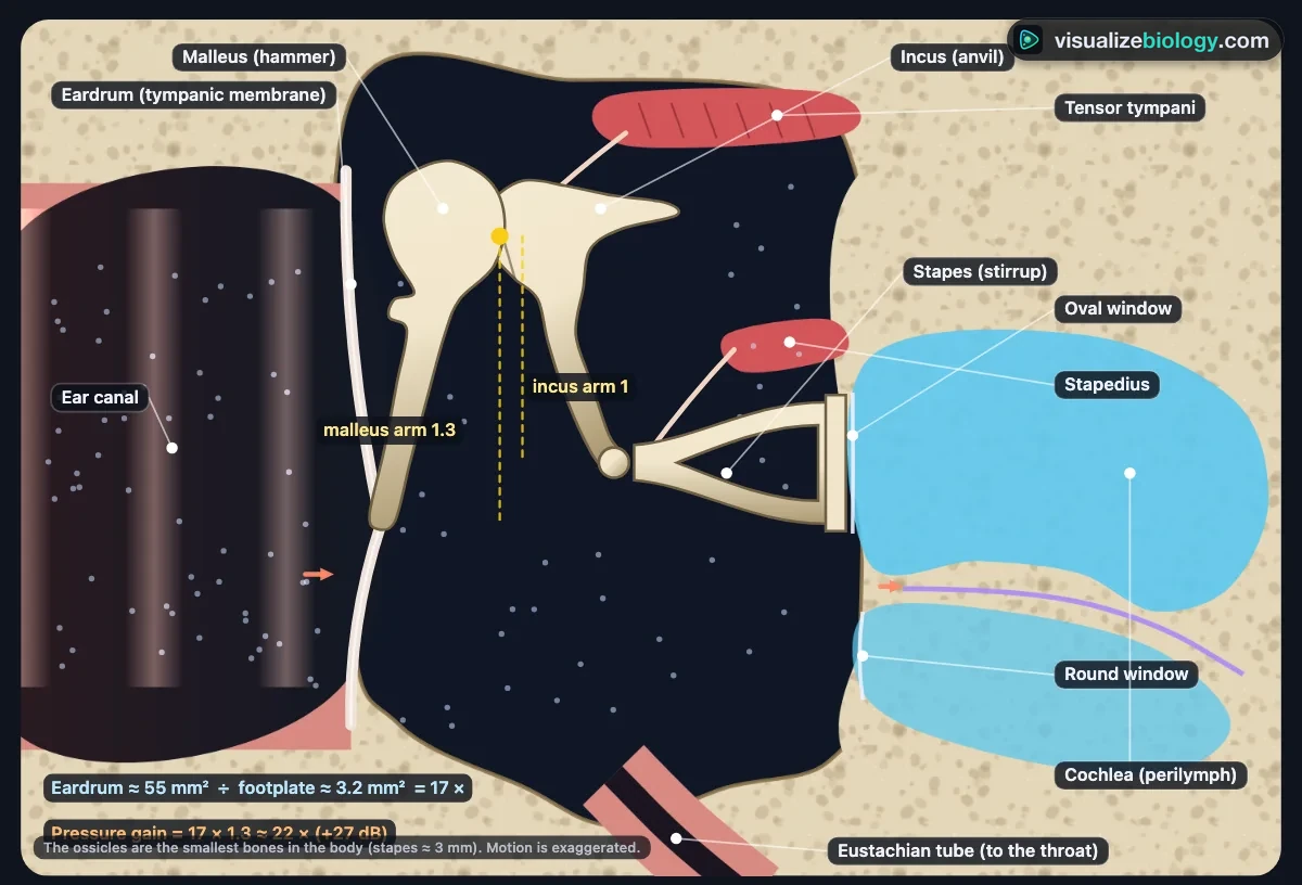
- Middle ear: the eardrum, malleus, incus and stapes close up, with the lever arms and areas that multiply the pressure about 22 times, the two muscles (the stapedius contracts above 85 dB) and the Eustachian tube.
- Cochlea: the cochlea unrolled. The stapes pushes the oval window, the round window bulges out, and a traveling wave runs along the basilar membrane and peaks at the place that matches the pitch. The scale shows the distance from the base and the frequency mapped there.
- Balance: the semicircular canals, utricle and saccule, with the cupula and an otolith organ close up and a head on a spinning chair.
- To the brain: the auditory pathway from both ears through the brainstem to the auditory cortex, and the direction of the sound.
3 Sound: pitch, loudness, direction
- In the Sound panel choose a pure tone or an everyday sound (whisper, conversation, traffic, a rock concert, a jet), set the Pitch from 20 Hz to 20 000 Hz and the Loudness in decibels, and move where the sound comes from.
- Hear it plays the tone through your speakers or headphones. Turn the volume down first. The loudness is not calibrated and loud settings are capped, so this is not a hearing test.
- Record this sound writes the sound and what the ear did with it in the lab notebook.
4 The ear's condition
The Ear panel (or the Ear menu in the command bar) changes the ear: a healthy teenager, age 70, noise damage, ear wax, glue ear, a perforated eardrum, otosclerosis, Ménière's disease, drug damage or profound deafness, which can be given a cochlear implant. Each one changes the picture, the audiogram and what is heard. These are educational models of typical cases, not medical advice.
5 Balance and flying
In the Move panel, Spin turns the chair at the chosen speed and Stop stops it: watch the cupula, the endolymph arrows and the Balance graph, and see the "feels like" turning carry on after the stop. Tilt head and Accelerate slide the crystals of the utricle. Take off and Land fly a plane (one second is fifteen seconds of flight): on the way up the ears pop by themselves; on the way down press Swallow or Valsalva to open the Eustachian tube.
6 Follow the sound
Press 🎧 on the transport bar to follow a sound step by step from the air to the auditory cortex. Each step picks the view and lights up the parts. Use ‹ and › to step, ❚❚ to pause and Done to finish.
7 Experiment mode and the lab report
Seven practicals: parts of the ear, an audiogram hearing test on a patient, the Rinne and Weber tuning fork tests, pitch and place in the cochlea, how the middle ear amplifies, sound localization, and spin and dizziness. Each shows its aim, what you need and a safety note; the method ticks off as you work, a conclusion question checks your result, and Create lab report builds a PDF with your results, pictures and calculations. The audiometer and the tuning fork are in the Lab panel.
8 Quiz and units
The practice quiz covers the parts of the ear, hearing, the middle ear, the cochlea, balance, sound and decibel calculations, hearing loss and tests, and the brain, each with a worked answer. Take the quiz gives five mixed questions and a star rating. SI | US switches lengths, speeds and pressures between metric (m, mm, m/s, kPa) and US units (ft, in, ft/s, psi). Decibels and hertz are the same in both.
9 Keyboard and gestures
- Space plays or stops the sound, 1–5 choose the view, ↑ and ↓ change the pitch by half an octave, L labels, F follow the sound, C, W, A, G the close-up, wave, audiogram and graph, S Status, Esc stops the journey.
- In 3D, drag to turn the ear and right-drag to move it; a plain scroll over it scrolls the page, so hold Ctrl or ⌘ (or click the ear first) to zoom. On a phone one finger scrolls the page and two fingers turn the ear.
- Right-click the ear or the close-up to save it as a picture or copy the readings.
How the human ear works: anatomy, hearing and balance
The ear does two jobs. It turns sound, tiny ripples of pressure in the air, into nerve impulses the brain hears as speech, music and noise, and it senses the head's position and movement so we keep our balance. It does both with the same kind of receptor: the hair cell. This simulator lets you watch a working ear: play a sound and follow it from the eardrum to the cochlea and the brain, or spin a chair and see why you feel dizzy.
- Subject
- The human ear: outer, middle and inner ear, the auditory nerve and pathway
- It shows
- Sound waves, eardrum and ossicles, the traveling wave in the cochlea, hair cells, balance, air pressure, hearing loss
- Views
- Whole ear, middle ear, cochlea unrolled, balance, to the brain; close-ups, wave, audiogram, graphs
- Modes
- Simulate (with Follow the sound), Experiment (7 practicals) and Quiz
- Price
- Free, no account or download; works on Chromebooks, tablets and phones
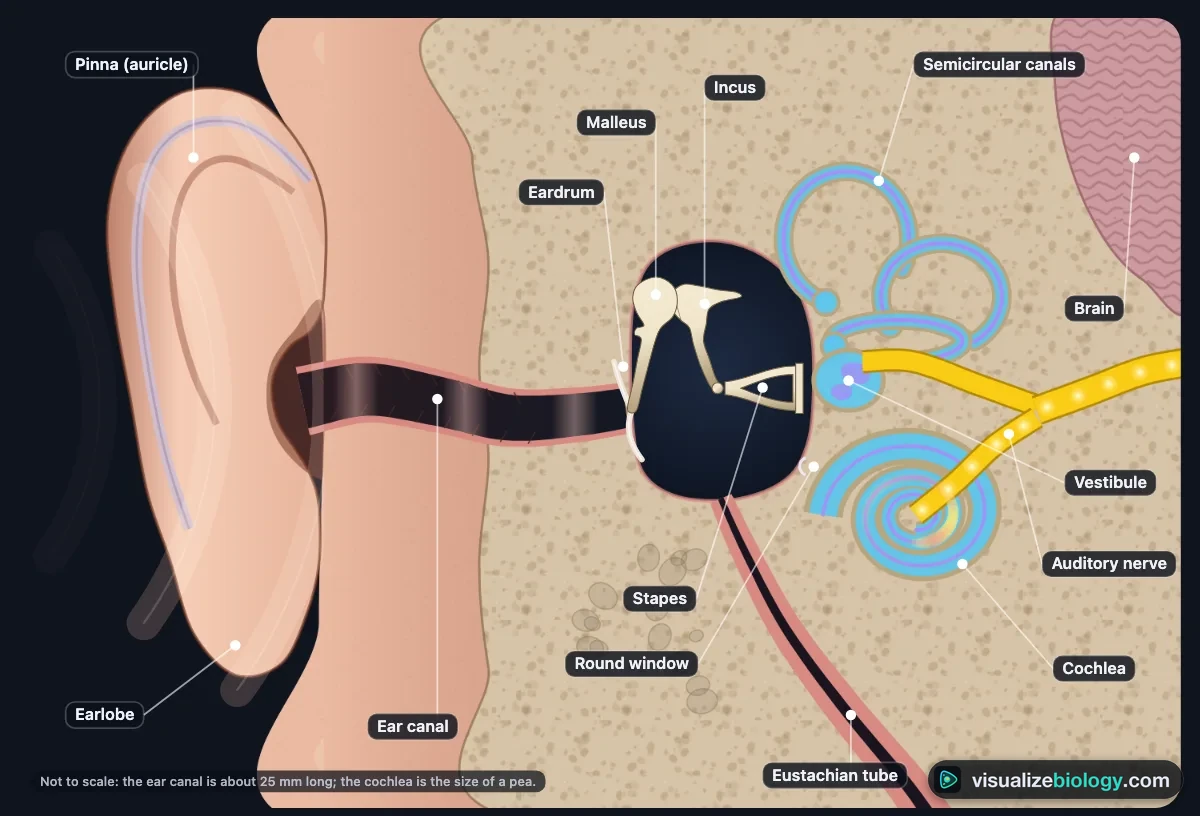
The parts of the ear and their functions
| Part | Where | What it does |
|---|---|---|
| Pinna (auricle) | Outer ear | Collects sound and funnels it into the canal; its folds help tell up from down and front from back |
| Ear canal | Outer ear | A 25 mm tube to the eardrum; wax and hairs trap dirt; it boosts pitches near 3000 Hz |
| Eardrum (tympanic membrane) | Between outer and middle ear | Vibrates with the sound and passes it to the malleus |
| Malleus, incus, stapes (ossicles) | Middle ear | Pass the vibration to the oval window and increase its pressure about 22 times |
| Eustachian (auditory) tube | Middle ear to throat | Keeps the air pressure equal on both sides of the eardrum |
| Oval and round windows | Wall of the inner ear | The stapes pushes the oval window; the round window bulges out to let the fluid move |
| Cochlea | Inner ear | Fluid-filled spiral where hair cells turn vibrations into nerve impulses, each pitch at its own place |
| Semicircular canals | Inner ear | Three fluid-filled loops that sense the head turning |
| Utricle and saccule | Inner ear (vestibule) | Sense gravity, tilt and straight-line movement |
| Auditory (vestibulocochlear) nerve | To the brain | Carries the impulses to the brainstem and the auditory cortex |
How we hear: from sound wave to nerve impulse
- Sound waves enter the ear canal and make the eardrum vibrate.
- The malleus, incus and stapes pass the vibration on and amplify it.
- The stapes pushes the oval window, making pressure waves in the fluid of the cochlea.
- A traveling wave runs along the basilar membrane and peaks at the place that matches the pitch.
- There, the hair cells of the organ of Corti bend against the tectorial membrane; ion channels open, K⁺ flows in and the cells release neurotransmitter.
- The auditory nerve fires impulses to the brainstem, which compares the two ears, and on to the auditory cortex in the temporal lobe.
Why the middle ear amplifies sound
Air is light and easy to move; the liquid in the cochlea is not. If sound went straight from air into liquid, about 99.9 % of its energy would bounce off. The middle ear solves this impedance mismatch in two ways: the eardrum is about 17 times the area of the stapes footplate, so the same force is squeezed onto a smaller area, and the ossicles are a lever that adds about 1.3 times. Together they raise the pressure about 22 times, or 27 decibels. Two tiny muscles, the tensor tympani and the stapedius, stiffen the chain when sounds are very loud: the acoustic reflex.
The cochlea and the place theory of pitch
Uncoiled, the cochlea is a 35 mm tube divided lengthways by the basilar membrane. Near the oval window (the base) the membrane is narrow and stiff; at the far end (the apex) it is five times wider and floppy. So each frequency makes it vibrate most at one place: about 20 000 Hz at the base, 1000 Hz about 21 mm along and 20 Hz at the apex. This tonotopic map is kept all the way to the auditory cortex, and it is why age and loud noise, which kill hair cells near the base first, take away high pitches first. Young people hear about 20 Hz to 20 000 Hz; the outer hair cells act as an amplifier that makes the quietest sounds audible and pitch sharp. At low pitches the nerve also fires in time with the wave (frequency theory, the volley principle).

Balance: the semicircular canals, utricle and saccule
The three semicircular canals lie at right angles to each other, so together they detect turning in any direction. When the head turns, the fluid inside (endolymph) lags behind and bends the cupula, a jelly flap over the hair cells in each ampulla. At a steady speed the fluid catches up and the feeling fades; stop suddenly and the fluid carries on, so you feel as if you are spinning the other way: that is why you are dizzy after a spin. The utricle and saccule carry tiny crystals (otoconia) that slide when the head tilts or speeds up in a straight line. When these signals disagree with the eyes, many people feel motion sickness.
Hearing loss: conductive vs sensorineural
| Feature | Conductive | Sensorineural |
|---|---|---|
| Where | Outer or middle ear | Cochlea (hair cells) or nerve |
| Examples | Wax, glue ear, perforated eardrum, otosclerosis | Aging, loud noise, some drugs, Ménière's disease |
| Audiogram | Bone conduction normal; air–bone gap | Air and bone equally reduced, often worse at high pitch |
| Rinne test | Negative on the bad side | Positive |
| Weber test | Heard in the bad ear | Heard in the better ear |
| Treatment | Often curable | Permanent: hearing aids, cochlear implants |
Loud sound is the cause you can prevent: 85 dB is safe for about 8 hours a day, and every 3 dB more halves the time, so earbuds at full volume (about 100 dB) are safe for only about 15 minutes.
Experiments you can do
| Practical | What it shows | Course links |
|---|---|---|
| Parts of the ear | The structures of the outer, middle and inner ear and their jobs | NGSS MS-LS1-8; IGCSE sense organs |
| Hearing test: an audiogram | Thresholds at each pitch; types and grades of hearing loss | HS anatomy & physiology; IB neurobiology |
| Rinne and Weber tests | Conductive vs sensorineural hearing loss | Anatomy & physiology; intro college |
| Pitch and place in the cochlea | Place theory: distance along the membrane against frequency | AP Psychology; IB; HS-LS1-2 |
| How the middle ear amplifies | Area ratio × lever = pressure gain | GCSE/IB physics of hearing |
| Sound localization | Interaural time difference against angle | AP Psychology; IB |
| Spin and dizziness | The semicircular canals and inertia of the endolymph | GCSE; HS-LS1-3 |
Who uses this simulator?
High school and early college students use it to see what a diagram cannot show: the eardrum moving, the ossicles rocking, the wave finding its place in the cochlea, hair cells firing and the fluid in the canals lagging behind a turn. Teachers project it to ask "what will happen if…" before pressing play (a higher pitch, a louder sound, wax in the canal, a stiff stapes), and set the practicals when there is no audiometer or tuning fork to hand. It is an educational model built from published physiology, not a hearing test or medical advice; it needs no login and runs on school Chromebooks.
Frequently asked questions
What are the parts of the ear and what do they do?
The ear has three parts. The outer ear (the pinna and the ear canal) collects sound and funnels it to the eardrum. The middle ear is an air-filled space holding the three smallest bones in the body, the malleus (hammer), incus (anvil) and stapes (stirrup); they pass the eardrum's vibration to the oval window of the inner ear and raise its pressure about 22 times. The Eustachian tube joins the middle ear to the throat and keeps the air pressure equal on both sides of the eardrum. The inner ear is filled with fluid: the snail-shaped cochlea turns vibrations into nerve impulses for hearing, and the vestibule and three semicircular canals sense balance. The auditory (vestibulocochlear) nerve carries the signals to the brain.
How does the ear turn sound into nerve impulses?
Sound waves make the eardrum vibrate. The malleus, incus and stapes pass the vibration on, and the stapes pushes the oval window in and out, making pressure waves in the fluid of the cochlea. These set up a traveling wave on the basilar membrane, which is narrow and stiff at the base and wide and floppy at the apex, so each pitch makes it vibrate most at one place: high pitches near the base, low pitches near the apex. Where it moves most, the hair cells of the organ of Corti rub against the tectorial membrane and their stereocilia bend. This opens ion channels, potassium ions flow in, the hair cell depolarizes and releases a neurotransmitter, and the auditory nerve fires impulses to the brainstem and the auditory cortex in the temporal lobe.
What is the difference between conductive and sensorineural hearing loss?
Conductive hearing loss means sound cannot get through the outer or middle ear: ear wax, fluid behind the eardrum (glue ear), a hole in the eardrum or a stiff stapes (otosclerosis). The cochlea still works, so bone conduction is normal and there is a gap between air and bone thresholds on the audiogram; it is often treatable. Sensorineural hearing loss means the cochlea's hair cells or the auditory nerve are damaged, for example by aging, loud noise or some drugs; air and bone conduction are equally reduced, high pitches usually go first, and the loss is permanent, so hearing aids or a cochlear implant are used. The Rinne test is negative and the Weber test lateralizes to the bad ear in a conductive loss, while in a sensorineural loss Rinne is positive and Weber lateralizes to the better ear.
Explore related simulators
See the malleus, incus and stapes in place inside the temporal bone, with the other 203 bones, in the Interactive 3D Skeleton. Compare the ear's pump-free fluid waves with a real pump in the Heart Simulator, where the heart sounds you hear through a stethoscope come from its valves. Look inside the animal cells that hair cells are specialized from in the Virtual Living Cell, see real tissue sections down the Virtual Microscope, and watch the water movement that keeps the inner ear's fluids in balance in the Osmosis Lab. Follow light instead of sound through the other sense organ in the head in the Human Eye Simulator, and see where the auditory pathway ends, in the temporal lobe, in the interactive 3D brain model.
Sources: OpenStax Biology 2e, section 36.4 (Hearing and Vestibular Sensation); OpenStax Anatomy and Physiology 2e, section 14.1; Urry et al., Campbell Biology 12th edition, chapter 50; Pickles, An Introduction to the Physiology of Hearing 4th edition; Greenwood (1990) J. Acoust. Soc. Am. 87:2592; WHO World Report on Hearing (2021); ISO 7029; NIOSH (1998). The model is an educational simplification of these sources; vibrations are slowed down and enlarged so they can be seen.
Updated