
Human Eye Simulator
Open up the eye in 2D and 3D, follow light from the cornea to the retina, watch the lens focus near and far and the pupil react to light, and see the world through eyes with myopia, cataract or color blindness.
Experiment
Sharp imageHealthy6 m
This eye
Focus—
Visual acuity—
Accommodation—
Eye's power—
Pupil—
Near point—
Far point—
Light—
Image on retina—
Show me
What the eye looks at
Light
Iris color
The eye
no lens
Lab
View
Hover or tap any part to name it. In 3D, drag to turn the eye; hold Ctrl or ⌘ and scroll (or click the eye first) to zoom.
What the eye is doing
Lab notebook
Your measurements are written here as you record them in a practical. Export a CSV, or turn the session into a PDF lab report.
| # | Subject | Conditions | Measurement | Result |
|---|
Concept library
Click "New question" to begin.
Click "Start quiz" for 5 questions on the parts of the eye, focusing, the retina, the pupil reflex, eye problems, color vision and calculations.
User Guide: Human Eye Simulator
1 Overview
Simulate shows a human eye looking at something: an eye chart, a book, a candle or a tree. On the left is the eye itself (a labeled section in 2D, or a 3D eyeball); on the right, What it sees shows the world through that eye. Experiment has seven practicals that end in a PDF lab report, and Quiz has a practice quiz with worked answers and a 5-question quiz. Everything comes from one model of the eye (a Le Grand schematic eye), so the rays, the picture and the numbers always agree. It is an educational model, not medical advice.
2 The 2D and 3D eye
- The eye opens in 3D on a computer or tablet and in 2D on a phone; the 2D | 3D switch changes it and remembers your choice.
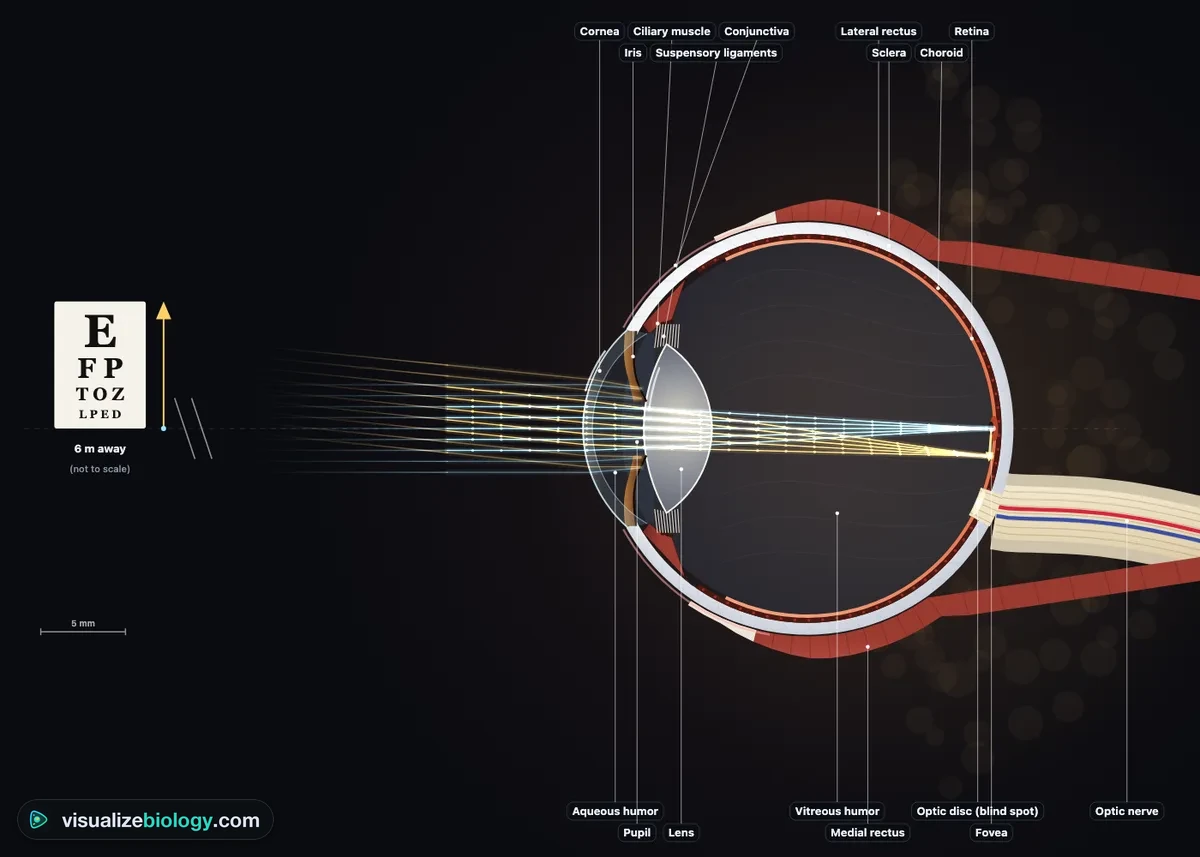
- In 2D, Section is a horizontal cut through the right eye seen from above: light comes in from the left, the temporal side is up and the nose side (with the optic nerve) is down. Front shows both eyes face-on, so you can watch both pupils.
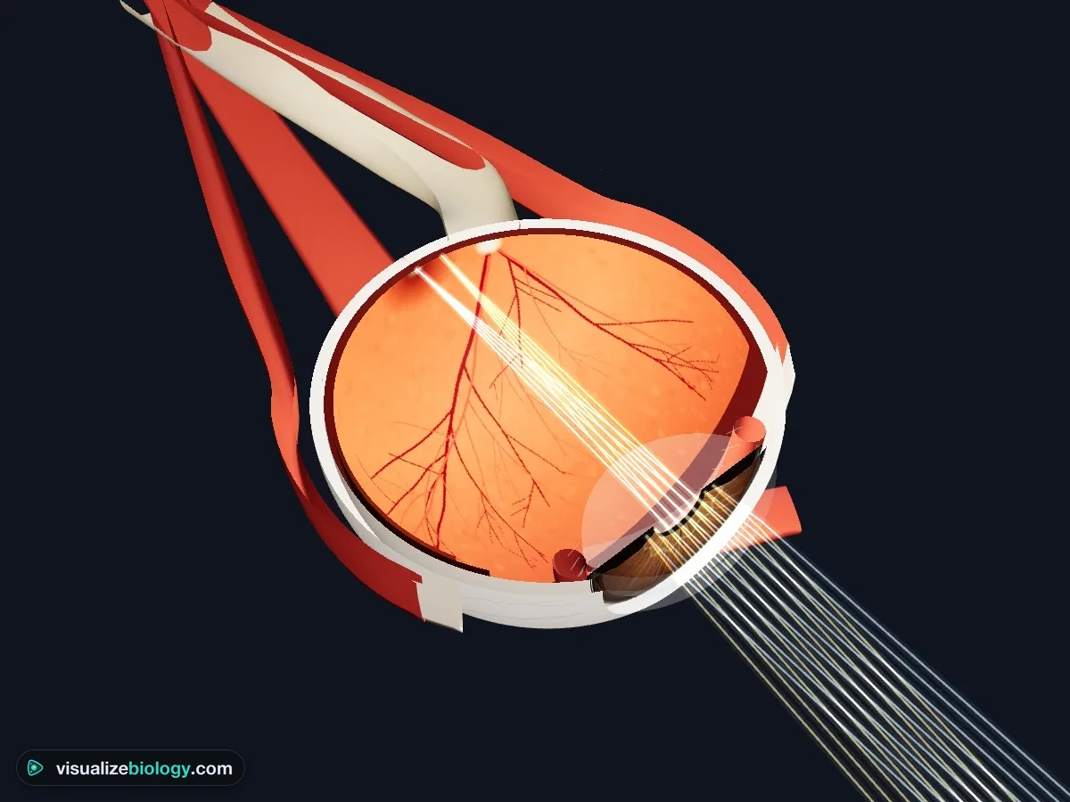
- In 3D, Outside shows the eyeball with its six muscles, Cutaway lifts the top half off along the same section, Retina looks at the back of the eye (the optic disc, macula and blood vessels) and Behind shows the optic nerve. Drag to turn it.
- Hover or tap any part to name it; click it to learn what it does. Labels and Light rays switch on and off in the corner.
3 Looking near and far
In the Object panel choose what the eye looks at and move it with the distance slider (or 25 cm, 1 m, 6 m, far away). Watch the lens: for near things the ciliary muscle contracts, the suspensory ligaments go slack and the lens rounds up (accommodation). Gold and blue lines are light rays from the top and the middle of the object: they cross and land upside down on the retina. If they meet in front of or behind the retina, the image is blurred and the focus point is marked. Look near / Look far in the action bar jumps between a book and the chart.
4 Light and the pupil
The Light panel goes from starlight to bright sun. The pupil follows in about half a second when the light gets brighter, and more slowly when it gets dimmer. 🔦 Shine a torch sends a flash into the right eye: in Front view both pupils shrink (the consensual reflex). In starlight the "What it sees" view loses its color, because only the rods work. You can also choose the color of the iris.
5 Eye problems and glasses
Choose a condition in the command bar: myopia, hyperopia, presbyopia, astigmatism, cataract, glaucoma, or one of three kinds of color blindness. In the Eye panel, change the age (the near point recedes as the lens stiffens), put Glasses on and change the lens power, or press 👓 Prescribe to choose the right lens: concave (minus) for myopia, convex (plus) for hyperopia and reading.
6 What it sees, retina, pupil, graphs
- What it sees: the object at its true size and blur for this eye, with its visual acuity (6/6 or 20/20 is normal).
- Retina: light enters from the top, passes the nerve cells and reaches the rods and cones; their signals run back through bipolar and ganglion cells into the optic nerve. Compare the fovea (cones, one to one) with the edge of the retina (rods, many to one), and try colored light.
- Pupil: the circular and radial muscles of the iris, and the reflex arc from the retina through the brain to the iris.
- Graph: accommodation against distance, pupil size against light, rods and cones across the retina, the three cones' sensitivity, dark adaptation, and the near point against age. Each graph marks this eye now.
7 "Show me" stories
Follow the light takes light from the object through the cornea, pupil, lens and vitreous to the retina and the optic nerve. How it focuses brings a book close and shows the ciliary muscle, ligaments and lens at work. Pupil reflex dims the light, then shines a torch into one eye. Use ‹ and › to step, ❚❚ to pause and Done to finish.
8 Experiment mode and the lab report
Seven practicals: find your blind spot, near point and age, the pupil reflex, prescribe glasses, test color vision, dark adaptation, and a cow's eye dissection. Each shows its aim, what you need and a safety note; the method ticks off as you work, a conclusion question checks your result, and Create lab report builds a PDF with your results and calculations. Labels, light rays, Prescribe and the stories are learning aids and are locked here.
9 Quiz, units and keyboard
- The practice quiz has eight topics, each with a worked answer; Take the quiz gives five mixed questions and a star rating.
- SI | US shows distances in centimeters and meters with acuity as 6/6, or in inches and feet with acuity as 20/20. The eye's own sizes (a 24 mm eyeball, a 3 mm pupil) and lens powers in dioptres are the same in both, as in every eye clinic.
- Keys: N look near/far, T torch, L labels, R rays, G prescribe glasses, 1–4 choose the right-hand view, S Status, Esc stops a story. A plain scroll over the 3D eye scrolls the page; hold Ctrl or ⌘ to zoom. Right-click a view to save it as a picture or copy the readings.
The human eye: its parts, how it works, and what goes wrong
Your eye works like a camera that focuses itself. Light from an object passes through the clear cornea, through the pupil in the colored iris, and through the lens, which together bend it to a sharp, upside-down image on the retina at the back. The retina's rods and cones turn the light into nerve impulses, and the optic nerve carries them to the brain, which turns the picture the right way up. This simulator lets you look inside the eye in 2D and 3D, trace the light, and see what the eye sees.
- Model
- A Le Grand schematic eye: about 24 mm long and 60 dioptres, the cornea 43 D and the lens 22 D
- It shows
- Light rays to the retina, accommodation, the pupil reflex, rods and cones, the blind spot, eye defects and their lenses
- Views
- 2D section and front view, a 3D eyeball you can cut open, what the eye sees, the retina, the iris and six graphs
- Modes
- Simulate (with guided stories), Experiment (7 practicals) and Quiz
- Price
- Free, no account or download; works on Chromebooks, tablets and phones
Parts of the eye and their functions
| Part | What it does |
|---|---|
| Cornea | Clear front window; does about two-thirds of the focusing |
| Iris | Colored ring of muscle; changes the size of the pupil |
| Pupil | The hole that lets light in |
| Lens | Fine-tunes the focus by changing shape |
| Ciliary muscle and suspensory ligaments | Change the shape of the lens |
| Aqueous and vitreous humor | Clear fluid and jelly that keep the eye in shape |
| Retina | Light-sensitive layer of rods and cones |
| Fovea | Pit packed with cones: the sharpest vision |
| Optic disc (blind spot) | Where the optic nerve leaves; no rods or cones |
| Optic nerve | Carries impulses to the brain |
| Choroid | Blood vessels that feed the retina; dark pigment stops reflections |
| Sclera | Tough white coat that protects the eye |
How does the eye focus? Accommodation
Light bends most where it enters the cornea, because the change from air to the eye is the biggest. The lens adds the fine adjustment. Light from a distant object arrives almost parallel and needs little bending: the ciliary muscle relaxes, the suspensory ligaments are pulled tight and the lens is stretched thin. Light from a near object spreads out more: the ciliary muscle contracts, the ligaments go slack and the elastic lens rounds up and gets fatter. This is accommodation. Lens power in dioptres is 1 ÷ focal length in meters, so focusing at 25 cm needs about 1 ÷ 0.25 = 4 D more. The closest point you can focus is the near point: about 7 cm at age 10, 25 cm by the mid-40s and over 1 m by 60, as the lens hardens (presbyopia).
Rods and cones
| Feature | Rods | Cones |
|---|---|---|
| Work in | Dim light | Bright light |
| Color | No: black and white | Yes: three kinds (blue, green, red) |
| Detail | Low: many share one nerve fiber | High: one to one in the fovea |
| Where | Edge of the retina; none in the fovea | Packed in the fovea |
| Number | About 92 million | About 4.6 million |
Light has to pass through the retina's nerve cells before it reaches the rods and cones. Where those nerve fibers leave the eye, at the optic disc, there are no receptors at all: the blind spot, about 15° to the side of where you look.
Find your blind spot: cover your left eye, look at the cross with your right eye, and move your face slowly towards the screen. At some distance the dot vanishes: its image has landed on your optic disc.
The pupil reflex
In bright light the circular muscles of the iris contract and the pupil gets smaller; in dim light the radial muscles contract and it gets bigger. It is a reflex arc: retina (receptor) → optic nerve (sensory neuron) → brain → nerve to the iris (motor neuron) → iris muscles (effector). Light in one eye makes both pupils shrink.
Myopia vs hyperopia, and other eye problems
| Problem | Cause | Image falls | Corrected by |
|---|---|---|---|
| Myopia (nearsighted) | Eyeball too long | In front of the retina | Concave (minus) lens |
| Hyperopia (farsighted) | Eyeball too short | Behind the retina | Convex (plus) lens |
| Presbyopia | Lens stiffens with age | Behind the retina (near things) | Convex reading lens |
| Astigmatism | Cornea curved unevenly | Two different distances | Cylindrical lens |
| Cataract | Cloudy lens | Scattered | Lens replaced |
Color vision and color blindness
Each color stimulates the three kinds of cone in its own mix. Without working red (L) or green (M) cones, reds and greens look alike: red–green color blindness, which is X-linked and affects about 8 % of men and 0.5 % of women. Missing blue (S) cones (tritanopia) is rare. Color-dot test plates hide a number in dots that differ only in a color the person cannot tell apart.
Experiments you can do
| Practical | What it shows | Course links |
|---|---|---|
| Find your blind spot | The optic disc has no receptors; it lies about 15° from the fovea | NGSS MS-LS1-8; AP Psychology |
| Near point and age | Accommodation weakens with age | AQA GCSE 4.5.2.3; NCERT Class 10 |
| Pupil reflex | A reflex arc, and both pupils respond | GCSE, IGCSE 0610 |
| Prescribe glasses | Concave lenses for myopia, convex for hyperopia | GCSE Biology and Physics |
| Color plates | Red–green and blue–yellow color blindness | A-level, AP Psychology |
| Dark adaptation | Cones adapt first, rods later (rod–cone break) | AQA A-level 3.6.1.2 |
| Cow's eye dissection | The parts of a mammal's eye, and the tapetum | Middle and high school |
Who uses this simulator?
Students use it to see what a diagram cannot show: the lens changing shape, the pupil reacting, the image landing upside down, and the world through other people's eyes. Teachers project it to ask "what happens if the eyeball is too long?" before pressing the button, and set the practicals when there is no time (or no cow's eyes) for the real thing. It is an educational model built from published optics and physiology; it needs no login and runs on school Chromebooks.
Frequently asked questions
What are the parts of the eye and their functions?
The cornea is the clear front window that does most of the focusing. The iris is the colored ring of muscle that changes the size of the pupil, the hole that lets light in. The lens, held by the suspensory ligaments and the ciliary muscle, fine-tunes the focus for near and far objects. The aqueous and vitreous humors fill the eye and keep its shape. The retina at the back contains the rods and cones that turn light into nerve impulses; the fovea gives the sharpest vision and the optic disc, where the optic nerve leaves, is the blind spot. The optic nerve carries the impulses to the brain. The choroid feeds the retina and absorbs stray light, and the tough white sclera protects the eye.
How does the eye focus on near and far objects?
By changing the shape of the lens, which is called accommodation. For a distant object the ciliary muscle relaxes, the suspensory ligaments are pulled tight and the lens is stretched thin, so it bends light only a little. For a near object the ciliary muscle contracts, the suspensory ligaments go slack and the elastic lens rounds up and gets fatter, so it bends light more. Either way the image lands sharp on the retina. With age the lens stiffens, the near point moves away and most people need reading glasses after about 45 (presbyopia).
What is the difference between rods and cones?
Rods work in dim light: they are very sensitive but see no color and little detail, because many rods share one nerve pathway to the brain. There are about 92 million, mostly around the edge of the retina. Cones work in bright light and give color and fine detail. There are about 4.6 million, of three kinds that respond best to blue, green and red light, packed most tightly in the fovea, where each cone has its own nerve pathway. That is why you see sharp colors when you look straight at something in daylight, but only grey shapes at night.
Explore related simulators
Open up another organ in 3D in the Heart Simulator. Use lenses the other way round, to magnify, in the Virtual Microscope. Look inside the cells the retina is made of in the Virtual Living Cell, see how the X chromosome passes on color blindness in the Meiosis Simulator, and explore the spectrum of light that plants use in the Photosynthesis Lab. Follow the other sense organ in the head, from sound wave to hair cell, in the Human Ear Simulator. Follow the signal from the retina to the visual cortex in the occipital lobe in the interactive 3D brain model. Compare the body's largest sense organ, with its receptors for touch, pain and temperature, in the Interactive Skin Diagram.
Sources: OpenStax Biology 2e, section 36.5 (Vision); OpenStax Anatomy and Physiology 2e, section 14.1; Urry et al., Campbell Biology 12th edition, chapter 50; Atchison & Smith, Optics of the Human Eye (the Le Grand eye); Curcio et al. 1990 (photoreceptor topography); Govardovskii et al. 2000 (visual pigment templates); de Groot & Gebhard 1952 (pupil size); Machado, Oliveira & Fernandes 2009 (color-vision deficiency); AQA GCSE Biology 8461, section 4.5.2.3. The simulator is an educational model, not medical advice: see an optometrist for any concern about your eyes.
Updated