
Interactive Skin Diagram: Layers of the Skin and How It Senses Touch
Cut open a block of skin to see the epidermis, dermis and hypodermis and every structure in them, then press, stroke and prick it to watch the touch receptors fire. See how skin keeps you at 37 °C and what UV light and heat do to it.
What you're looking at
Lab notebook
Every region you open, every touch, temperature and exposure is written here. Export a CSV, or turn the session into a PDF lab report.
| # | Skin | What | Result | Value |
|---|
Concept library
Click "New problem" to begin.
Click "Start quiz" for 5 questions on the layers of the skin, the epidermis, glands, touch receptors, temperature control and sun protection.
1 Overview
Simulate shows a block of skin cut open, on the left, in five views: Layers (every structure, labelled), Touch (make the receptors fire), Temperature (how skin keeps the body at 37 °C), Sun & burns (UV light, vitamin D, sunburn and how deep a burn goes) and Epidermis (its five strata, cell by cell). On the right, Nerve signals shows the impulses from eight kinds of receptor, the Body map picks a region of skin, and Graph plots the numbers. Experiment has five practicals that end in a PDF lab report, and Quiz has a practice quiz with worked solutions and a 5-question quiz.
2 Exploring the layers
- Tap any part of the section to see what it is, its job and a fact in What you're looking at; the camera zooms in during the tour. Hover (on a computer) for its name.
- Press ▶ in Layers for a guided tour from the stratum corneum down to the fat.
- In the Skin panel choose where on the body the skin comes from (fingertip, palm, sole, lip, cheek, forearm, back, scalp) and its type (Fitzpatrick I to VI). True proportions (or 📏 True scale) eases each layer to its real thickness: textbooks draw the epidermis far thicker than it is.
3 Touch and the nerve signals
- In the Touch view, press and hold on the skin's surface with the mouse or a finger (or press 👆 Touch). Choose the probe in the Touch panel: press, vibrate (set the frequency and amplitude), brush, stretch, pin, hot or cold.
- Receptors glow as they fire and their impulses run along the nerve (slowed down so you can follow them). Nerve signals shows every receptor's impulses over the last 4 seconds and what the brain feels.
- ▶ in Touch plays a demo of every kind of touch. Switch on Hear the nerve impulses in View, then tap a row of Nerve signals to choose which receptor you hear.
4 Temperature, sun and burns
In Temperature, set the air temperature, humidity and activity in the Climate panel and press ▶: the arterioles widen or narrow, sweat appears, hairs stand up and muscles shiver as the body's model needs. The Graph shows core and skin temperature, sweat and skin blood flow. In Sun & burns, press ☀️ to go into the sun and ▶ to run the clock; set the UV index and sunscreen in the Sun panel and your skin type in Skin. Switch to Hot burn to touch a hot surface and see the burn reach the epidermis, the dermis or right through it. Teach speed runs 2 minutes a second; 5 and 20 min/s are faster.
5 Experiment mode and the lab report
Five practicals: label the skin, the two-point touch test on five parts of the body, fast and slow receptors, keeping 37 °C in cold and hot air, and how long until sunburn. Each shows its aim, what you need and a real-lab safety note; the method ticks off as you work, a conclusion question checks your result, and Create lab report builds a PDF. The controls for each practical are in the Lab panel.
6 Units, keyboard and gestures
- SI | US switches between °C and millimetres and °F and inches (thousandths of an inch, mils, for layers under a millimetre). Cells stay in µm in both.
- Space plays or pauses, T does the view's main action, 1–5 choose the views, S opens Status and Esc clears the selection.
- A plain scroll over the section scrolls the page. Right-click the section or the graph to save it as an image.
Layers of the skin: an interactive labeled skin diagram
The skin is the largest organ of the body and the main part of the integumentary system. In an adult it covers about 1.8 m² and makes up around 16 % of body weight. It is built in layers, each with its own job: a thin outer epidermis, a thick dermis beneath it, and the fatty hypodermis that anchors it to the muscle. This interactive skin diagram draws the labeled cross-section every textbook uses and lets you take it apart, then makes it work: its receptors fire when you touch it, its glands and blood vessels respond to heat and cold, and its melanin meets the sun.
- Shows
- Epidermis (five strata), dermis (papillary and reticular), hypodermis and 25 structures you can click
- Regions
- Fingertip, palm, sole, lip, cheek, forearm, back and scalp: thick and thin skin
- Simulates
- Eight sensory receptors, temperature control, UV and vitamin D, sunburn and burn depth
- Modes
- Simulate, Experiment (5 practicals with a PDF report) and Quiz
- Price
- Free, with no account or download; works on Chromebooks, tablets and phones
Skin diagram labeled: every part and its function
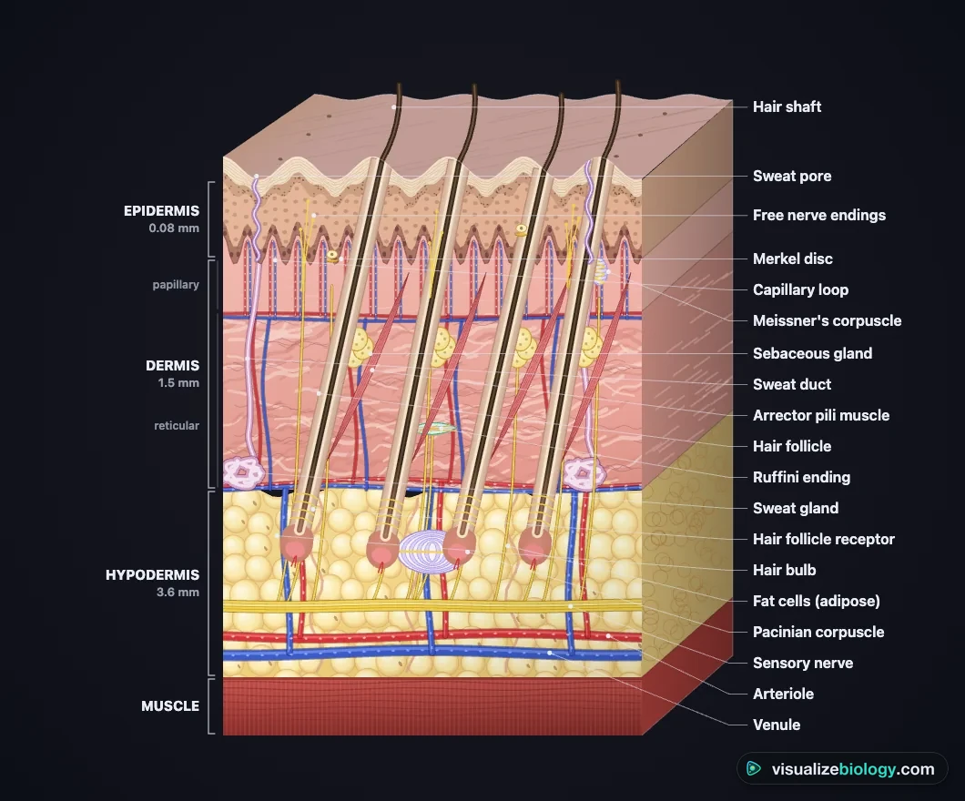
A labeled skin diagram shows a block of skin cut open, with the three layers bracketed on one side and the structures in each named on the other. These are the parts students are asked to label, from the surface down. Switch the labels off in the simulator and its Label the skin practical becomes a skin labeling quiz.
| Part | Layer | Function |
|---|---|---|
| Hair shaft | Above the epidermis | Insulation and protection; moving it is felt by the follicle's nerve endings |
| Sweat pore | Epidermis (surface) | Lets sweat out to evaporate and cool the body |
| Stratum corneum | Epidermis | Dead keratin-filled cells: the waterproof barrier |
| Stratum basale (melanocytes) | Epidermis | Makes new cells; melanin shields them from UV |
| Free nerve endings | Epidermis | Pain, heat, cold and itch |
| Merkel disc | Base of the epidermis | Steady pressure, edges and shapes |
| Dermal papilla and capillary loop | Papillary dermis | Feeds the epidermis; grips it; gives off heat |
| Meissner's corpuscle | Papillary dermis | Light touch and flutter |
| Sebaceous gland | Dermis | Sebum (oil) for supple, waterproof skin and hair |
| Arrector pili muscle | Dermis | Raises the hair: goosebumps |
| Hair follicle and hair bulb | Dermis and hypodermis | Grows the hair; its stem cells repair wounds |
| Eccrine sweat gland and duct | Deep dermis | Makes sweat for cooling |
| Ruffini ending | Reticular dermis | Skin stretch |
| Collagen and elastic fibres | Dermis | Strength and stretch |
| Pacinian corpuscle | Deep dermis and hypodermis | Vibration and deep pressure |
| Sensory nerve | Hypodermis | Carries impulses to the spinal cord and brain |
| Arteriole and venule | Hypodermis to dermis | Bring and drain blood; widen or narrow to control heat loss |
| Adipose (fat) cells | Hypodermis | Insulation, energy store, cushioning |
The 3 layers of the skin
| Layer | Made of | Contains | Main jobs |
|---|---|---|---|
| Epidermis (0.05–1.5 mm) | Stratified squamous epithelium, mostly keratinocytes; no blood vessels | Melanocytes, Langerhans cells, Merkel cells, free nerve endings | Waterproof barrier, protection from microbes and UV, vitamin D |
| Dermis (1–4 mm) | Connective tissue: collagen and elastic fibres made by fibroblasts | Blood vessels, nerves, touch receptors, sweat and sebaceous glands, hair follicles, arrector pili muscles | Strength and stretch, feeding the epidermis, sensation, temperature control |
| Hypodermis (subcutaneous layer) | Loose connective tissue and fat (adipose tissue) | Fat lobules, larger vessels and nerves, Pacinian corpuscles | Insulation, energy store, cushioning, anchors skin to muscle |
The dermis has two parts. The thin papillary dermis folds up into dermal papillae, each with a capillary loop that feeds the epidermis by diffusion, and in hairless skin a Meissner's corpuscle. The thick reticular dermis below is dense irregular connective tissue that gives skin its strength. Switch on True proportions in the simulator to see how thin the epidermis really is: on the forearm only about 0.08 mm, under 2 % of the depth to the muscle.
The 5 layers of the epidermis
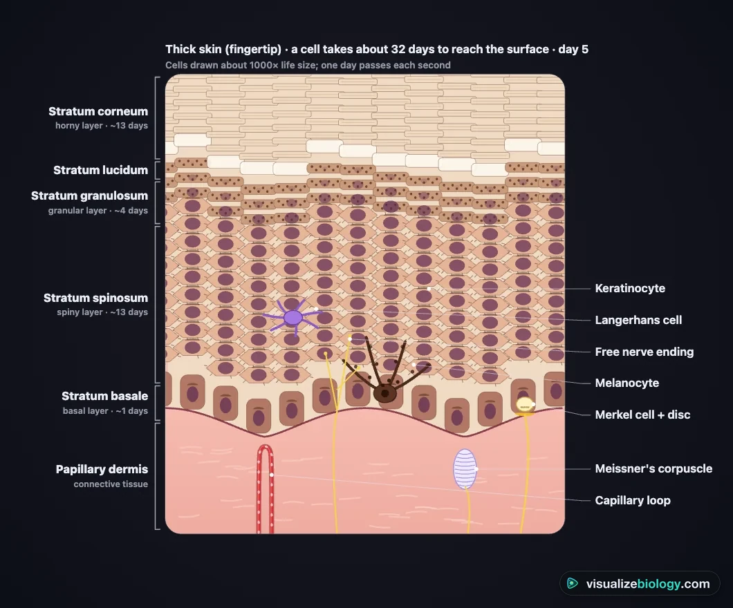
New cells are made by mitosis in the deepest layer and pushed up. As each keratinocyte rises it flattens, fills with the tough protein keratin, dies and is finally shed, about four weeks after it was born. The layers (strata) are named for what the cells look like on the way:
| Stratum (surface first) | What you see | What happens there |
|---|---|---|
| Corneum | 15–30 layers of flat, dead cells | The barrier; cells flake off (desquamation) |
| Lucidum | A clear band, thick skin only | Extra protection on palms and soles |
| Granulosum | Flattened cells with dark granules | Keratin forms, lipids seal the skin, cells die |
| Spinosum | Many-sided cells joined by desmosomes | Strength; Langerhans cells watch for microbes |
| Basale | One row of column-shaped stem cells | Cell division; melanocytes and Merkel cells |
A memory aid from the surface down is Come, Let's Get Sun Burned. Thick skin on the palms, soles and fingertips has all five layers, a very thick stratum corneum, friction ridges (fingerprints) and no hair. Thin skin covers the rest of the body: it has four layers, with hair follicles and sebaceous glands.
Sensory receptors in the skin: how we feel touch
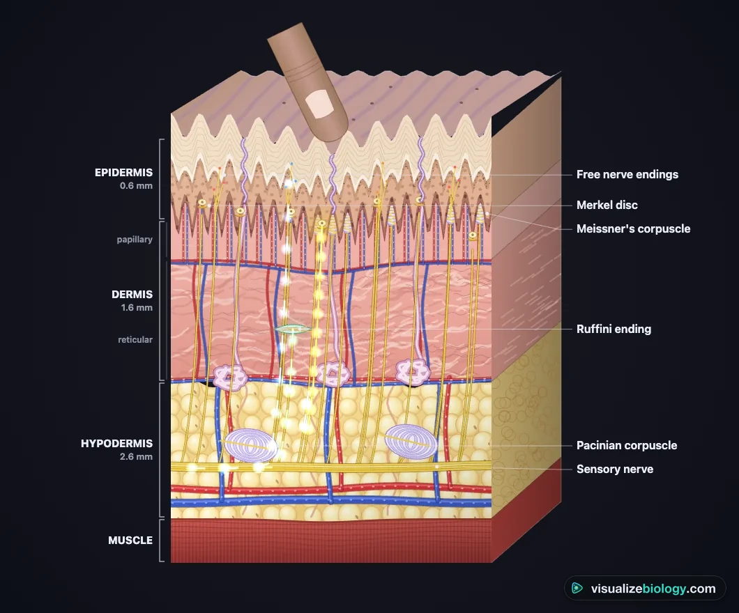
The skin is a sense organ. Each kind of receptor responds to one kind of stimulus and sends nerve impulses along sensory neurones to the spinal cord and the brain's sensory cortex. Some are rapidly adapting: they fire only when a stimulus starts or changes. Others are slowly adapting and keep firing as long as it lasts.
| Receptor | Where | Detects | Adapts |
|---|---|---|---|
| Meissner's corpuscle | Dermal papillae of hairless skin | Light touch, flutter (10–50 Hz), slip | Rapidly |
| Merkel disc | Base of the epidermis | Steady pressure, edges, shapes, Braille | Slowly |
| Pacinian (lamellar) corpuscle | Deep dermis and hypodermis | Vibration (best near 250 Hz), deep pressure | Very rapidly |
| Ruffini ending | Reticular dermis | Stretch, finger and joint position | Slowly |
| Hair follicle receptor | Round the hair follicle | Hairs moving | Rapidly |
| Free nerve endings | Into the epidermis | Pain, heat, cold, itch | Mostly slowly |
Fingertips have about 140 Meissner's corpuscles and 70 Merkel discs per cm², each with a receptive field a few millimetres across, so two points just 2–3 mm apart feel like two. On the back two points 4 cm apart can feel like one: this is two-point discrimination, and you can measure it in the Experiment mode.
Functions of the skin
- Protection: a barrier against microbes, chemicals, injury and UV light, and against losing water.
- Temperature regulation: sweating and vasodilation lose heat; vasoconstriction, goosebumps and fat keep it in.
- Sensation: touch, pressure, vibration, stretch, heat, cold, pain and itch.
- Vitamin D: UVB light turns 7-dehydrocholesterol in the epidermis into vitamin D₃.
- Excretion and storage: sweat removes a little salt and urea; the hypodermis stores fat.
How the skin controls body temperature
The hypothalamus keeps the core near 37 °C, and the skin is where most of the heat is lost or kept. When you are hot, arterioles in the dermis vasodilate, so more warm blood flows near the surface, and eccrine glands make sweat that cools you as it evaporates (about 2.4 kJ per gram). When you are cold, the arterioles vasoconstrict, the arrector pili muscles raise goosebumps, and the muscles shiver to make heat. In the simulator these follow Gagge's two-node model of the body: in 40 °C air, sweating is the only way left to lose heat.
Skin, UV light and burns
UVC is stopped by the ozone layer. UVB is absorbed in the epidermis: it makes vitamin D but also causes sunburn and damages DNA in the dividing basal cells. UVA reaches the dermis and breaks down collagen, ageing the skin. Melanin, made by melanocytes and passed to keratinocytes, absorbs both, so darker skin types take several times the dose to burn. Burns are graded by depth: first degree (epidermis only, red and painful), second degree (into the dermis, with blisters) and third degree (through the whole dermis, white or charred and painless because the nerve endings are destroyed). The Sun & burns view is an educational model, not medical advice.
Practicals and course links
| Practical | What you find out | Course links |
|---|---|---|
| Label the skin | The layers and structures on a skin section | IGCSE 14.4; A&P integumentary system; NCERT 11 |
| Two-point touch test | Which parts of the body feel finest touch, and why | NGSS MS-LS1-8; AP Psychology sensation; GCSE nervous system |
| Fast and slow receptors | Which receptors adapt to a steady press | AQA A-level 3.6.1.2 (Pacinian corpuscle) |
| Keeping 37 °C | How skin and core temperature change with the air | IGCSE 14.4; GCSE homeostasis; NGSS HS-LS1-3 |
| How long until sunburn? | How skin type, UV index and SPF change burning time | Health education; NGSS HS-LS1-2 |
Who uses this skin model?
Middle and high school students learning the structure of the skin, the sense of touch and homeostasis for NGSS, GCSE, IGCSE, A-level, IB and NCERT/CBSE courses (including NEET preparation), anatomy and physiology students meeting the integumentary system, and teachers who want a free, no-login labeled skin diagram (or a skin labeling quiz) on a classroom screen or students' Chromebooks.
Frequently asked questions
What are the 3 layers of the skin?
The skin has two layers, the epidermis and the dermis, sitting on a third, the hypodermis (subcutaneous layer). The epidermis is the thin outer layer of stratified squamous epithelium with no blood vessels; it makes the waterproof barrier and contains melanocytes. The dermis below is thick connective tissue of collagen and elastic fibres with blood vessels, nerves, touch receptors, sweat and sebaceous glands and hair follicles. The hypodermis is mostly fat: it insulates, stores energy and anchors the skin to the muscle underneath.
What are the 5 layers of the epidermis?
From the surface down: the stratum corneum (flat, dead cells full of keratin), the stratum lucidum (a clear layer found only in the thick skin of the palms and soles), the stratum granulosum (cells with dark keratohyalin granules that seal the skin and die), the stratum spinosum (many-sided cells joined by desmosomes, with Langerhans cells) and the stratum basale (one row of dividing stem cells with melanocytes and Merkel cells). A memory aid is Come, Let's Get Sun Burned. Thin skin has only four layers, without the stratum lucidum.
Which receptors in the skin detect touch?
Four kinds of mechanoreceptor. Meissner's corpuscles in the dermal papillae detect light touch and flutter and adapt quickly. Merkel discs at the base of the epidermis detect steady pressure, edges and shapes and adapt slowly. Pacinian corpuscles deep in the dermis and hypodermis detect vibration and deep pressure. Ruffini endings in the dermis detect stretch. Hair follicle receptors feel hairs moving, and free nerve endings detect pain, heat and cold.
What is the function of each layer of the skin?
The epidermis is the protective barrier: it keeps water in and microbes and UV light out, renews itself about every four weeks and makes melanin and vitamin D. The dermis gives the skin strength and stretch and carries the blood vessels, nerves, touch receptors, sweat and sebaceous glands and hair follicles, so it feeds the epidermis, senses touch, pain and temperature and helps control body temperature. The hypodermis is mostly fat: it insulates the body, stores energy, cushions it and anchors the skin to the muscle below.
What does a labeled skin diagram show?
A labeled skin diagram is a cross-section of a block of skin. It brackets the three layers (epidermis, dermis and hypodermis) and names the structures in them: the hair shaft, follicle and bulb, the sebaceous gland, the arrector pili muscle, the sweat gland, duct and pore, blood vessels and capillary loops, the sensory nerve and the touch receptors (Meissner's and Pacinian corpuscles, Merkel discs, Ruffini endings and free nerve endings), and fat cells. In thick skin it also shows the five strata of the epidermis.
Explore related simulators
Follow touch signals to the sensory cortex and its homunculus in the Interactive 3D Brain Model, and compare the skin with the body's other sense organs in the Human Eye Simulator and the Human Ear Simulator. See how the hypothalamus and hormones keep the body in balance in Hormone Quest: The Interactive Endocrine System Lab, and watch the cells of the stratum basale divide in the Mitosis Simulator. Look at a real skin section down the Virtual Microscope.
Sources: OpenStax Anatomy and Physiology 2e, chapter 5 (The Integumentary System); OpenStax Biology 2e, chapter 36.2 (Somatosensation); Johansson and Vallbo, Journal of Physiology 286 (1979), for receptor densities; Weinstein (1968), for two-point thresholds; Bolanowski et al., JASA 84 (1988), for vibration tuning; Gagge, Stolwijk and Nishi, ASHRAE Transactions 77 (1971), for temperature control; Henriques (1947), for burns; Fitzpatrick (1988), for skin types. Thicknesses and receptor counts outside the hand are scaled estimates; the simulation is an educational model, not medical advice.
Updated