
Virtual Fetal Pig Dissection: Pig Anatomy and the Fetal Circulation, Step by Step
Tie a fetal pig to the tray, cut it open with the scissors and lift out its organs one layer at a time, naming each one as you go. Then watch its heart, gut and kidneys at work in a see-through pig, and see how its blood changes course at birth.
What you're looking at
Lab notebook
Every step of your dissection, each cut's accuracy, each organ lifted out and every measurement is written here. Export a CSV, or turn the session into a PDF lab report.
| # | Specimen | What | Result | Value |
|---|
Concept library
Click "New problem" to begin.
Click "Start quiz" for 5 questions on the fetal pig's external features, mouth, organs, digestion, the fetal circulation, excretion and reproduction, glands and calculations.
1 Overview
Simulate puts a fetal pig on a dissecting tray, on the left, in four views: External (from above and below), Mouth (cut open at the jaw), Dissection and Systems (a see-through pig). The card on the right shows an Organ page, animated close-ups (How it works) and Graphs. Experiment has five practicals with a PDF lab report; Quiz has a practice quiz and a 5-question quiz.
2 External features and the mouth
- Tap any part to read what it is, its job and an exam fact in What you're looking at; hover (on a computer) for its name. ▶ plays a guided tour.
- ↻ Flip over turns the pig to see its belly: the umbilical cord, the teats and the clues to its sex.
- Choose the specimen size and sex in the Pig panel.
3 Doing the dissection
- Pick 🧵 String (the right instrument is picked for you at each step) and tap the four yellow rings at the hooves.
- With ✂️ Scissors, press on the yellow dot and drag along the yellow line. Each cut is graded.
- With 🤏 Forceps, tap the belly to fold the flaps back.
- Cut the umbilical vein, then up the thorax beside the sternum, across the top, and up the neck.
- Tap organs to name them; tap them with the forceps (or use the Organs panel) to lift them into the dish, top layer first.
✂️ Cut for me does the next step; ▶ Watch it done performs the whole dissection.
4 Systems, the fetal circulation and close-ups
Pick a system in the Systems panel. In the circulatory system every vessel is coloured by how much oxygen it carries; switch between Before birth and After birth to see the umbilical vessels and the fetal short cuts close. Tap any organ to open its Organ page: a labelled close-up, its parts, what it does, exam points and how it compares with a human.
5 Experiment mode and the lab report
Five practicals: external features and sexing, the mouth and pharynx, the full dissection (graded), age and gut length, and the fetal circulation. Each shows its aim, what you need and a real-lab safety note. Labels and "do it for me" are off. Finish with the conclusion question, then press 📄 Lab report (PDF).
6 Units, keyboard and gestures
- SI | US switches lengths between millimetres/centimetres and inches.
- Space plays or pauses, T does the view's next step, 1–4 pick the view, S opens the live readings, Esc clears the selection.
- Right-click the tray or the close-up to save an image or copy the details.
Virtual fetal pig dissection: mammal anatomy, step by step
The fetal pig is the standard dissection in American high school and college biology, because a pig is a mammal built like us: a four-chambered heart, lungs worked by a diaphragm, a liver, a stomach and a long small intestine, kidneys and a brain with twelve pairs of cranial nerves. The specimens are fetuses taken from sows at meat-packing plants; they never breathed or ate, which makes them a window on the fetal circulation too. This free virtual dissection follows the steps of a real lab guide, grades every cut you make, and lets you watch each organ system work, so it serves as preparation for the real thing or as the alternative many states let students choose.
- Specimen
- Fetal pig (Sus scrofa domesticus), small, regular or large (about 87 days to term); male or female
- You do
- Tie it down, cut the abdomen, thorax and neck, lift out organs with forceps, measure with a ruler
- Shows
- External anatomy, the mouth, 60+ organs and structures, 7 organ systems, the fetal circulation before and after birth
- Modes
- Simulate, Experiment (5 practicals with a PDF report) and Quiz
- Price
- Free, with no account or download; works on Chromebooks, tablets and phones
Fetal pig dissection steps
- External anatomy. Look at the pig from above and below, find the umbilical cord and teats, and decide its sex.
- The mouth. Cut the corners of the mouth back through the angles of the jaw and press the lower jaw down.
- Tie it down. Lay the pig on its back and tie a string round each leg, under the tray.
- Abdominal incisions. Lifting the wall as you cut, cut forward from the umbilical cord to the ribs, back on both sides of the cord, along the lower ribs and in front of the hind legs. Fold the flaps back.
- The cord flap. The umbilical vein runs from the cord to the liver and holds the flap down: cut it and fold the flap back with the bladder and umbilical arteries.
- Thorax and neck. Cut through the rib cartilage just beside the sternum (not the bone), across the top of the thorax, then up the neck to the chin.
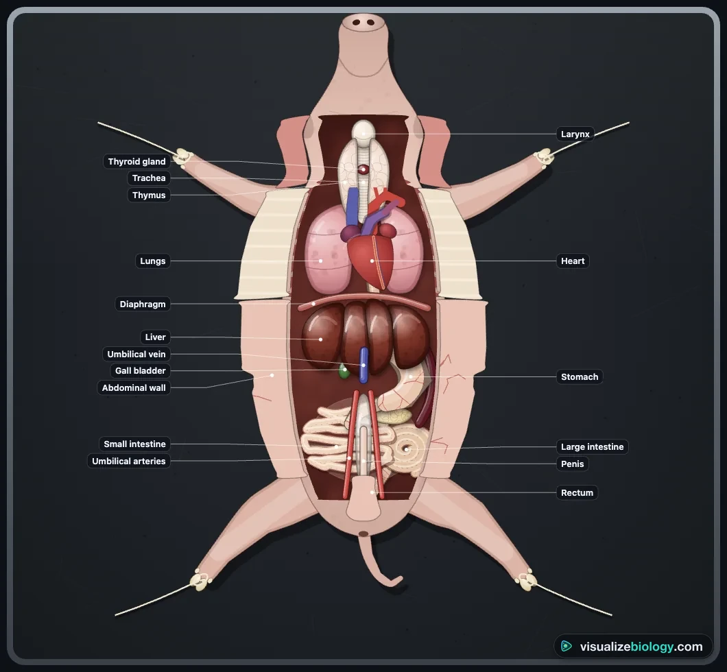
- Organs. Identify them in place, then lift out the thymus, heart, lungs, liver and digestive tract to see the kidneys, reproductive organs and great vessels on the back wall.
Fetal pig external anatomy and how to sex it
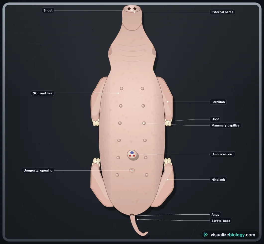
A pig has a head, neck, trunk and tail, a flat snout for rooting, outer ears and eyes usually still closed. Each foot has two main toes in hooves and two dewclaws: pigs are even-toed ungulates. On the belly are the cut umbilical cord and two rows of teats, which both sexes have. A male has a urogenital opening just behind the cord and scrotal sacs below the anus; a female has a small urogenital papilla just below the anus.
Inside the fetal pig's mouth
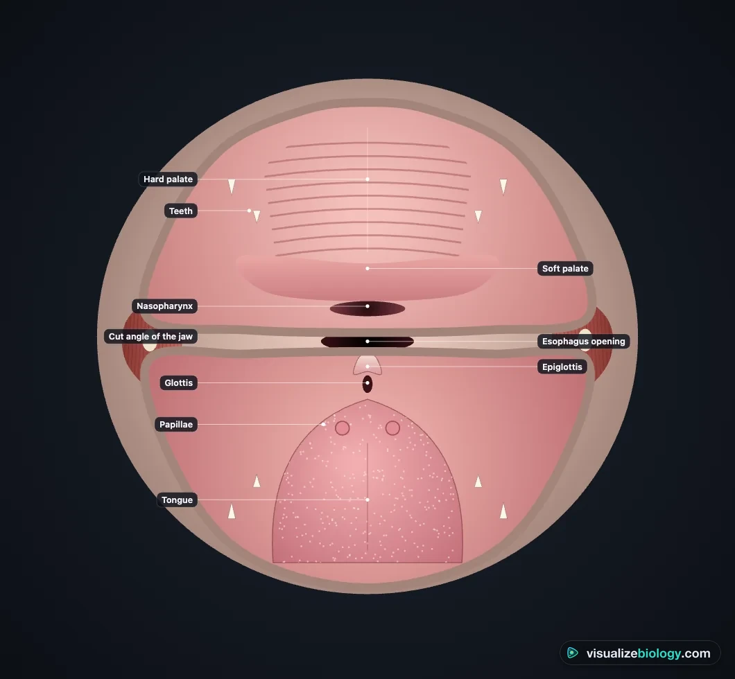
The roof has a ridged hard palate and a soft palate behind it, which separate the mouth from the nose so a piglet can suckle and breathe at once. Near term, eight sharp needle teeth have cut the gum. At the back, the epiglottis covers the glottis (the way to the larynx and trachea) when the pig swallows; behind it opens the esophagus, and above the soft palate the nasopharynx carries air from the nose.
Fetal pig internal anatomy: organs and their functions
| Organ | Where you find it | Function |
|---|---|---|
| Thymus and thyroid | Neck, beside and on the trachea | Trains T cells; thyroxine controls metabolism |
| Heart | Thorax, between the lungs, in the pericardium | Four chambers pump blood round the body and (after birth) the lungs |
| Lungs and diaphragm | Thorax; the diaphragm separates thorax from abdomen | Gas exchange after birth; the diaphragm pulls air in |
| Liver and gall bladder | Just behind the diaphragm | Makes bile (stored in the gall bladder); fetal blood cells |
| Stomach and spleen | The pig's left side, under the liver | Stores and digests food; the spleen filters blood |
| Small intestine and pancreas | Coiled in the middle, held by the mesentery | Digests and absorbs food; enzymes and insulin |
| Spiral colon and rectum | Lower left; back to the anus | Absorbs water; stores meconium in the fetus |
| Kidneys, ureters, bladder | Back wall; bladder between the umbilical arteries | Make, carry and store urine |
| Testes or ovaries and uterus | Scrotum; or behind the kidneys | Make sperm or eggs; a two-horned uterus holds a litter |
The fetal circulation and what changes at birth
A fetus breathes through its placenta. Blood about 80 % saturated with oxygen arrives in the single umbilical vein; the ductus venosus sends much of it past the liver; the foramen ovale lets it cross between the atria; and the ductus arteriosus sends most of the right ventricle's output into the aorta instead of the fluid-filled lungs, which get only about a tenth of the blood. Two umbilical arteries return oxygen-poor blood to the placenta. At birth the cord is cut and the lungs fill with air: the shunts close and the heart becomes a double pump like ours. Compare it with the adult cardiac cycle in the Heart Simulator: Blood Flow & Cardiac Cycle, and follow how a fetus forms in the Human Embryo Development Simulator.
Male vs female fetal pig
| Male | Female | |
|---|---|---|
| Outside | Urogenital opening just behind the cord; scrotal sacs | Urogenital papilla below the anus |
| Gonads | Testes in the scrotum or inguinal canals | Small ovaries behind the kidneys |
| Ducts | Vas deferens loops over the ureter to the urethra; penis under the belly skin | Oviducts, two uterine horns, uterine body, vagina |
How old is a fetal pig?
Pig gestation lasts about 114 days. Measure the crown–rump length (top of the head between the ears to the base of the tail) and use age ≈ 3 × length (cm) + 21 days: a 27 cm pig is about 102 days old. The small intestine of a near-term fetus is about ten times its body length.
Practicals and course links
| Practical | What you find out | Course links |
|---|---|---|
| External features and sexing | Mammal features; male or female | High school biology; anatomy and physiology |
| Mouth and pharynx | Palates, epiglottis, teeth | Fetal pig lab guides |
| The full dissection | How the organs are arranged in neck, thorax and abdomen | NGSS HS-LS1-2; AP Biology |
| Age and gut length | Fetal age; intestine vs body length | High school biology |
| Fetal circulation | Shunts, oxygen and the changes at birth | AP Biology; anatomy and physiology |
Who uses this virtual fetal pig dissection?
High school students doing the fetal pig lab or its alternative, AP Biology and anatomy and physiology students revising mammalian organ systems, pre-nursing and pre-vet students, and teachers who want a free, no-login dissection that runs on Chromebooks. It is an educational model, not a substitute for veterinary or medical training.
Frequently asked questions
How do you tell if a fetal pig is male or female?
Look at the belly near the hind legs. A male has a urogenital opening on the belly just behind the umbilical cord (the end of the penis) and two scrotal sacs below the anus. A female has no opening behind the cord; instead a small fleshy urogenital papilla sits just below the anus. Do not use the teats: male and female pigs both have two rows of them.
How old is a fetal pig used in dissection?
Pig gestation is about 114 days. A fetal pig's age can be estimated from its crown–rump length (from the top of the head between the ears to the base of the tail): age in days ≈ 3 × length in cm + 21. School specimens are usually 23–33 cm long, so they are about 90 to 114 days old, a few weeks before birth or at term.
Why does the umbilical vein carry oxygen-rich blood?
Before birth the fetus gets its oxygen from the placenta, not its lungs. The single umbilical vein carries blood about 80 % saturated with oxygen from the placenta through the cord to the liver and heart; the two umbilical arteries carry oxygen-poor blood back. Veins are named for the direction of flow (towards the heart), not for their oxygen.
What are the steps of a fetal pig dissection?
Study the external features and sex the pig, then cut the angles of the jaw to look inside the mouth. Tie the pig on its back by its legs. Cut the abdominal wall forward from the umbilical cord to the ribs, back on both sides of the cord, along the lower ribs and in front of the hind legs, and fold the flaps back. Cut the umbilical vein to free the cord flap, open the thorax through the rib cartilage beside the sternum, and open the neck to the chin. Identify the organs in place, then lift out the thymus, heart, lungs, liver and digestive tract to see the kidneys, reproductive organs and great vessels on the back wall.
Explore related simulators
Compare a mammal with an amphibian in the Virtual Frog Dissection, and the pig's organs with ours in the Human Digestive System Simulator and the Respiratory System Simulator.
Sources: OpenStax Biology 2e, chapters 34, 39, 40 and 43; Marrable, The Embryonic Pig (1971); Rudolph, Circulation Research 57 (1985); Getty (ed.), Sisson and Grossman's The Anatomy of the Domestic Animals (1975). An educational model.
Updated