
Human Embryo Development Simulator: From Fertilization to Birth in 3D
Follow one human life from the moment a sperm meets the egg, through all 23 Carnegie stages of the embryo and the fetal months, to birth, in 2D or 3D and at its real size. Watch it divide, implant, fold into a body, grow a beating heart, limbs and a face, then grow from 3 cm to 36 cm, and tap any part to name it.
What's happening
Lab notebook
Every milestone (first cleavage, implantation, the first heartbeat, limb buds…) is written here with the day it happened. Export a CSV, or turn the session into a PDF lab report.
| # | Age | What | Result | Value |
|---|
Concept library
Click "New problem" to begin.
Click "Start quiz" for 5 questions: the first week, implantation and germ layers, the organs, timing and dating, and a calculation.
1 Overview
Simulate shows one human conceptus on the left, in 2D or 3D, and close-ups on the right: the Timeline of organs and critical periods, Inside cross-sections, Growth graphs and the Mother. One model runs everything on one clock in days after fertilization, from day 0 to day 60, through the 23 Carnegie stages. Experiment has five practicals that end in a PDF lab report, and Quiz has a practice quiz with worked solutions and a 5-question quiz.
2 Playing through development
- Press ▶ (or Space) to play. At Teach speed every stage and fetal period gets a few seconds on screen, so fertilization to birth takes about three minutes; 1 h/s is good for watching the first cell divisions, 1 d/s for the embryo, 1 wk/s for the fetal months. › jumps to the next stage or period. The speed, labels, yolk sac, 3D cut-away and sound are in the toolbar at the top of the embryo view.
- The strip under the clock has one segment per Carnegie stage. The clock shows days after fertilization; Status also shows the age of the pregnancy (2 weeks more).
- Under the embryo view, the Journey panel goes straight to any Carnegie stage or fetal period, or to a moment (hatching, the first heartbeat…). Tap a stage on the Timeline to go there.
3 Views, labels and parts
- The view buttons at the top left change with the stage: Whole or Section for the first week, the implantation section, Top or Section for the embryonic disc, and Side, Front or Back for the embryo from week 4.
- The camera fits the subject: a 0.15 mm zygote is magnified hundreds of times, a 30 mm embryo a few times. The scale bar and "× life size" keep the real size honest.
- Tap any part to name it and see what it becomes. In 3D drag to turn the embryo; hold Ctrl or ⌘ and scroll (or pinch) to zoom; switch on 3D cut-away to open the blastocyst or the disc.
4 The close-ups
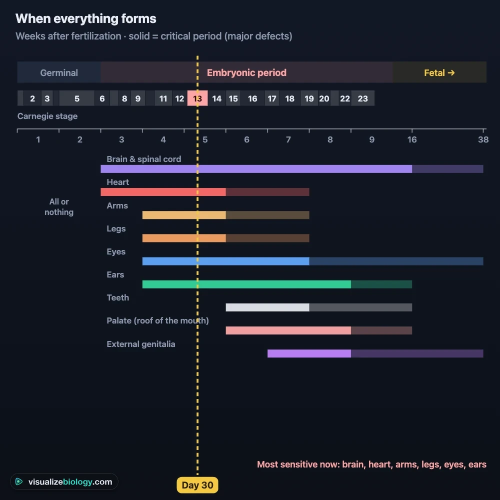
Timeline: the 23 stages over weeks 1–8 and when each organ forms, with its critical period. Inside: fertilization step by step, gastrulation and the germ layers, neurulation, the heart, the brain vesicles and the hand, each following today's stage. Growth: length, somites, heart rate, cell number in week 1 and fetal weight to birth. Mother: where the conceptus is in the uterine tube or uterus, and the pregnancy calendar.
5 Experiment mode and the lab report
Five practicals: stage the embryo, time the first divisions, count somites to find the age, measure the crown–rump length with calipers to date a pregnancy, and count the heartbeat across weeks. Each shows its aim, what you need and a safety or ethics note; the method ticks off as you work, a conclusion question checks your result, and Create lab report builds a PDF.
6 Units and keyboard
- SI | US switches the embryo's length between millimetres and inches and the fetal weight between grams and ounces or pounds. Cells and the disc stay in µm and mm in both.
- Space plays or pauses, → jumps to the next stage, ← to the previous one, S opens Status and Esc clears the selection. Right-click the embryo or the close-up to save it as an image.
Human embryo development, stage by stage
In the 8 weeks after fertilization a single cell becomes an embryo about 3 cm long with every major organ in place. This human embryo development simulator follows that whole embryonic period on one clock, through all 23 Carnegie stages, in 2D or 3D. Each stage is drawn at its real size from the measurements embryologists use, with its key features, and every structure can be tapped and named.
- Covers
- Fertilization to birth (day 0–266): all 23 Carnegie stages and the fetal months
- Views
- 2D textbook drawings and a 3D embryo; cross-sections, growth graphs, the mother
- Data
- Ages and lengths from O'Rahilly and Müller (2010); cleavage times from time-lapse IVF studies
- Modes
- Simulate, Experiment (5 practicals with a PDF report) and Quiz
- Price
- Free, with no account or download; works on Chromebooks, tablets and phones
Stages of embryonic development, week by week
| Week | What happens | Size |
|---|---|---|
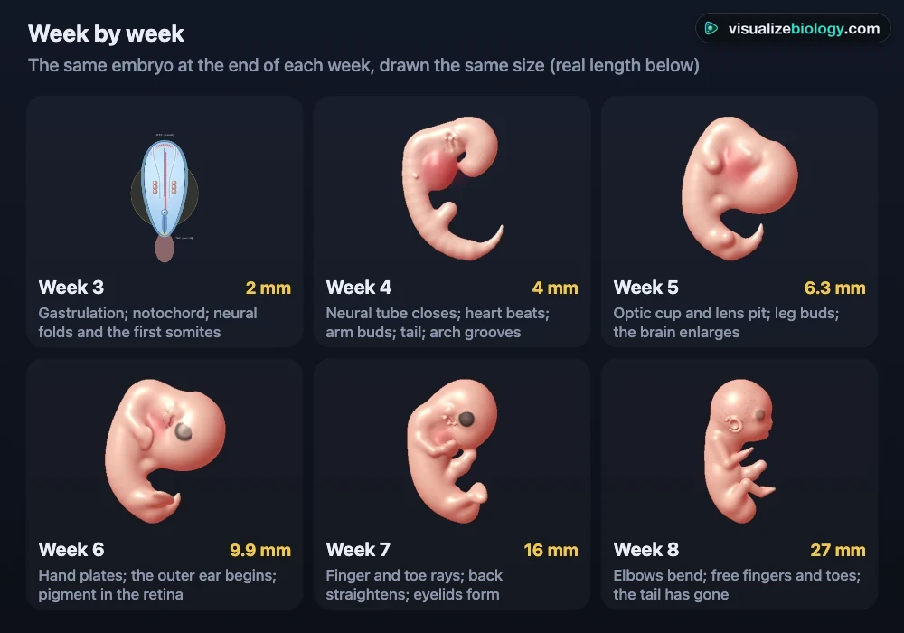
| 1 | Fertilization in the uterine tube; cleavage to 2, 4 and 8 cells; the morula enters the uterus (day 3–4); the blastocyst hatches and attaches (day 5–7) | 0.1–0.2 mm |
| 2 | Implantation; a two-layered disc (epiblast and hypoblast) between the amniotic cavity and the yolk sac; the placenta starts and makes hCG | 0.1–0.2 mm disc |
| 3 | The primitive streak and gastrulation: three germ layers; the notochord and the neural plate; the first somites | 0.4–2.5 mm |
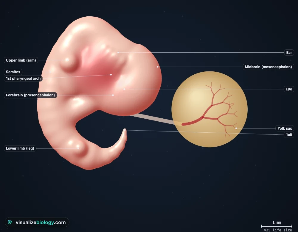
| 4 | The neural tube closes (day 22–28); the heart beats from day 22; the embryo folds into a C; arm and leg buds | 2.5–6 mm |
| 5 | Lens and optic cup; hand plate; five brain vesicles | 5–9 mm |
| 6 | Foot plate; retinal pigment; the outer ear begins; the gut grows into the cord | 8–14 mm |
| 7 | Finger and toe rays; elbows; eyelids; bone begins to form | 13–18 mm |
| 8 | Fingers and toes free; the tail has gone; the head is rounded | 18–31 mm |
The 23 Carnegie stages
Because embryos of the same age differ, embryologists stage them by their features, not their age: the number of cells or somites, the limb buds, the hand and foot plates, the digits, the eye and the ear. The Carnegie system, named after the Carnegie Institution's collection, has 23 stages up to day 60. In the simulator the Journey panel goes to any of them.
| Stage | Days | Length | Key feature |
|---|---|---|---|
| 1 | 1 | 0.1–0.15 mm | Zygote |
| 2–3 | 2–6 | 0.1–0.2 mm | Cleavage, morula, free blastocyst |
| 4–5 | 6–13 | 0.1–0.2 mm | Attachment and implantation; bilaminar disc |
| 6–8 | 13–19 | 0.2–1.5 mm | Primitive streak, gastrulation, neural plate |
| 9–12 | 19–28 | 1.5–5 mm | Somites, neural tube closure, heartbeat, arm buds |
| 13–16 | 28–42 | 4–11 mm | Leg buds, lens, hand plate, foot plate, retinal pigment |
| 17–20 | 42–53 | 11–22 mm | Finger and toe rays, elbows, scalp vessels |
| 21–23 | 53–60 | 22–31 mm | Free fingers and toes, eyelids, no tail |
From fertilization to the blastocyst
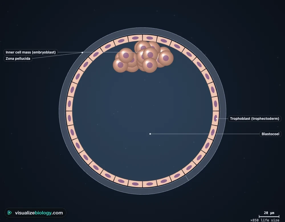
A sperm digests a path through the zona pellucida with its acrosome and fuses with the oocyte, which hardens its zona so no other sperm can enter and finishes meiosis. The two pronuclei meet and the zygote divides about 26 hours later. The cells (blastomeres) halve in size at each division, so the embryo stays about 0.1 mm across. At 8 cells they compact; at 16 or more they form a morula, which reaches the uterus on day 3–4. Fluid then gathers inside: the blastocyst has an outer trophoblast, which becomes the placenta, and an inner cell mass, which becomes the embryo. It hatches from its zona and implants in the lining of the uterus from day 6–7.
Implantation and the three germ layers
In week 2 the embryo sinks into the endometrium, and its syncytiotrophoblast opens the mother's capillaries and makes hCG, the hormone a pregnancy test detects. The inner cell mass becomes a two-layered disc between the amniotic cavity and the yolk sac. In week 3 a groove, the primitive streak, forms at the tail end, and epiblast cells pour through it: this is gastrulation, which makes the three germ layers. Ectoderm becomes the nervous system and skin, mesoderm the heart, blood, muscle and bone, and endoderm the lining of the gut and lungs.
Weeks 4–8: the organs form
The neural plate rolls into the neural tube, which closes from the neck toward both ends between days 22 and 28; if it does not, the result is spina bifida or anencephaly, which folic acid helps prevent. The heart tube starts beating about day 22. The flat disc folds into a C-shaped body, somites line the back, pharyngeal arches build the face and neck, and limb buds grow into hands and feet whose fingers and toes separate by programmed cell death. To see how the heart works once it has formed, open the Heart & Cardiac Cycle simulator.
Fetal development, month by month
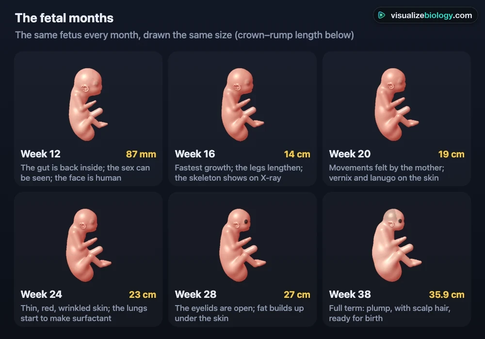
From week 9 the organs are all in place and the fetus mainly grows: its crown–rump length goes from about 5 cm to 36 cm and its weight from a few grams to about 3.4 kg. The head, half the length at week 9, becomes about a third of it by birth as the trunk and legs catch up. In the simulator the Journey panel has eight fetal periods after the 23 Carnegie stages, and the Timeline shows the fetal months side by side.
| Weeks after fertilization | Crown–rump length | Weight | What happens |
|---|---|---|---|
| 9–12 | 5–9 cm | 8–45 g | Eyelids fuse; the gut returns from the cord; the sex can be seen by week 12 |
| 13–16 | 9–14 cm | 45–200 g | Fastest growth; bones ossify; the legs lengthen |
| 17–20 | 14–19 cm | 200–460 g | The mother feels movements; vernix and lanugo cover the skin |
| 21–25 | 19–24 cm | 460–900 g | Thin, red, wrinkled skin; the lungs start to make surfactant |
| 26–29 | 24–27 cm | 0.9–1.5 kg | The eyelids open; fat builds up under the skin |
| 30–38 | 27–36 cm | 1.5–3.4 kg | The fetus fills out, gains about 30 g a day and is ready for birth |
Embryo vs fetus
From fertilization to the end of week 8 the developing human is an embryo; from week 9 to birth it is a fetus. The embryonic period is when the organs form; the fetal period is mostly growth and maturation, from about 3 cm and 3 g to about 50 cm and 3.4 kg. Doctors count pregnancy from the first day of the last period, about 2 weeks before fertilization, so week 8 of the embryo is week 10 of the pregnancy.
Critical periods and teratogens
Each organ is most easily damaged by drugs, infections or radiation while it forms. In weeks 1–2 an insult usually either ends the pregnancy or does no harm; weeks 3–8 are the most sensitive, when major malformations can result (thalidomide caused limb defects when taken between about days 20 and 36). The simulator's Timeline shows each organ's critical period. It is an educational model, not medical advice.
Practicals and course links
| Practical | What you find out | Course links |
|---|---|---|
| Stage the embryo | The features that define each Carnegie stage | AP Biology; IB D3.1; A&P |
| Time the first divisions | How fast cleavage doubles the cells | NGSS HS-LS1-4; NCERT 12 |
| Count somites, find the age | Somites as a developmental clock | AP Biology; college embryology |
| Measure the crown–rump length | How an ultrasound dates a pregnancy | Health science; A&P |
| Heart rate across weeks | How the embryonic heart rate rises | NGSS HS-LS1-2; health science |
Who uses this embryo development simulator?
High school students learning human reproduction and development (AP Biology, IB, GCSE and NCERT Class 12), college students in anatomy and embryology who need to picture the Carnegie stages in 3D, nursing and health-science students learning how a pregnancy is dated, and teachers who want a free, no-login embryo development simulation for a classroom screen or Chromebooks. The cells it starts from are the subject of the Mitosis Simulator; the gametes that make the zygote come from meiosis.
Frequently asked questions
What are the stages of embryonic development?
Fertilization makes a zygote; cleavage divides it into a ball of cells, the morula, by day 3–4; the blastocyst forms on day 4–5 and implants in the uterus from day 6–7. In week 2 it becomes a two-layered disc, and in week 3 gastrulation makes three germ layers (ectoderm, mesoderm and endoderm). Weeks 3 to 8 are organogenesis: the neural tube, heart, limbs, eyes and ears form. Embryologists divide these first 8 weeks into 23 Carnegie stages, from stage 1 (the zygote) to stage 23 (about day 56–60), after which the embryo is called a fetus.
How big is a human embryo at 8 weeks?
At the end of the embryonic period, 8 weeks after fertilization (10 weeks after the last menstrual period), the embryo is about 27–31 mm long from crown to rump and weighs about 2–3 g. It has a rounded head, eyelids that nearly cover the eyes, arms bent at the elbows, free fingers and toes, and no tail. At 4 weeks it is only about 4–5 mm long, and at 2 weeks the embryonic disc is about 0.2 mm across.
When does an embryo's heart start beating?
The heart tube starts to beat about 22 days after fertilization (Carnegie stage 10), when the embryo is about 2–3 mm long; it is the first organ to work. On ultrasound a heartbeat is usually seen from about 6 weeks of pregnancy counted from the last period. The rate rises from about 80–110 beats a minute in week 4–5 to about 170–175 in week 7–8, then settles toward 140–160.
Explore related simulators
See how a cell copies itself before each cleavage in the Mitosis Simulator, and how the egg and sperm get half the chromosomes in the Meiosis Simulator. The heart that starts beating on day 22 is pumping blood like the adult heart in the Heart & Cardiac Cycle simulator. Compare a very different development, complete metamorphosis, in the Butterfly Life Cycle Simulator, and see the cells of the blood first made in the yolk sac in the Interactive Blood Cell Model.
Sources: O'Rahilly and Müller (2010), Cells Tissues Organs 192, for the Carnegie stages, ages and lengths; Moore, Persaud and Torchia, The Developing Human, 11th edition, for the critical periods; Sadler, Langman's Medical Embryology, 14th edition; Meseguer et al. (2011), Human Reproduction 26, for the cleavage times; Robinson and Fleming (1975), BJOG 82, for dating by crown–rump length; Doubilet and Benson (1995), Journal of Ultrasound in Medicine 14, for the embryonic heart rate; OpenStax Biology 2e, chapter 43. Ages of human embryos are estimates and every embryo differs; the simulator is an educational model and not medical advice.
Updated