
Plant Tissues Virtual Lab: Stem and Root Cross Sections Under the Microscope
Put a sunflower stem, a maize stem, a buttercup root and a maize root under the microscope. Stain them, zoom from ×40 to ×400, click any tissue to see what its cells look like and do, draw the low-power plan, and see at a glance how monocot and dicot stems and roots differ.
About this tissue
Lab notebook
Every slide you put on the stage, every stain and every measurement is written here. Export a CSV, or turn the session into a PDF lab report.
| # | Slide | What | Result | Value |
|---|
Concept library
Click "New problem" to begin.
Click "Start quiz" for 5 questions on plant tissues, stems, roots, stains and microscope skills.
1 Overview
Simulate puts a prepared slide under a compound microscope on the left: the circle of light shows the stained section, inverted as in a real microscope, with labels naming the tissues in view and a slide map in the corner. The Plan view shows the same section as a low-power plan diagram. On the right, the close-ups show the tissue you picked, the monocot and dicot versions side by side, every tissue under every stain, and graphs.
2 Using the microscope
- Pick ×40 to see the whole section, ×100 for the tissues and ×400 for single cells (or press + and −, or hold Ctrl/⌘ and scroll).
- Drag the slide to move it, or click the slide map; the arrow keys move it too and C centres it.
- Measure (M): drag across something; the length is written in the notebook. Graticule (G) puts the eyepiece scale in the field.
- Fine focus blurs the section if you move it away from 0: the section is only about 15 µm thick.
3 Slides, stains and tissues
- Slide: a dicot stem (sunflower), a monocot stem (maize), a dicot root (buttercup) and a monocot root (maize).
- Stain: safranin + fast green (lignin red, cellulose green), toluidine blue (lignin blue-green, cellulose purple), phloroglucinol + HCl (lignin red), iodine (starch blue-black) or unstained.
- Click any tissue, label or chip to see it in the Tissue close-up and in "About this tissue".
4 Experiment mode and the lab report
Six practicals: identify four mystery sections, draw and label a plan, test for lignin and starch, count the vascular bundles, measure xylem vessels and count the xylem groups of two roots. Each ticks off its method as you work and ends with a conclusion question; "Lab report (PDF)" turns your notebook into a report.
5 Units and keyboard
- SI | US shows cell sizes in µm or mil (1 mil = 25.4 µm) and section sizes in mm or inches; magnifications have no unit.
- 1 Microscope, 2 Plan, + − objective, arrows move the slide, M measure, G graticule, L labels, C centre, Esc clears.
A plant tissues virtual lab for stems and roots
Prepared slides of a sunflower stem, a maize stem, a buttercup root and a maize root are the classic way to learn plant tissues, and the classic way to get confused: a cross section is a wheel of red and green cells, and it takes practice to see the tissues in it. This virtual lab puts the four slides under a compound microscope at ×40, ×100 and ×400. Every cell is drawn at its real size from one model of the section, so the labels, the plan diagram and the close-ups always agree with what you see. Click any tissue to see its cells cut across and along, with its job and the point examiners look for.
- Slides
- Sunflower stem, maize stem, buttercup root, maize root, in transverse section
- Stains
- Safranin + fast green, toluidine blue, phloroglucinol + HCl, iodine, unstained
- Courses
- IGCSE and GCSE transport in plants; A-level, IB and AP plant tissues; NCERT Class 11 anatomy of flowering plants
- Modes
- Simulate, Experiment (6 practicals with a PDF report) and Quiz
- Price
- Free, with no account or download; works on Chromebooks, tablets and phones
Types of plant tissues
New cells come from meristems, such as the cambium. The permanent tissues fall into three systems: dermal (the epidermis, with a cuticle on a stem and root hairs on a root), ground (parenchyma, collenchyma and sclerenchyma) and vascular (xylem and phloem). A simple tissue is made of one kind of cell; a complex tissue of several.
| Tissue | Cells and walls | Living? | Job |
|---|---|---|---|
| Parenchyma | Large, thin cellulose walls, air spaces between them | Yes | Storage, photosynthesis in green cortex |
| Collenchyma | Corners thickened with cellulose and pectin | Yes | Flexible support in young stems |
| Sclerenchyma | Thick lignified walls, tiny lumen | No | Rigid support (fibres) |
| Xylem (complex) | Vessels, tracheids, fibres; lignified | Mostly no | Water and mineral ions up; support |
| Phloem (complex) | Sieve tubes with sieve plates, companion cells | Yes | Sugars and amino acids, up or down |
Dicot stem cross section
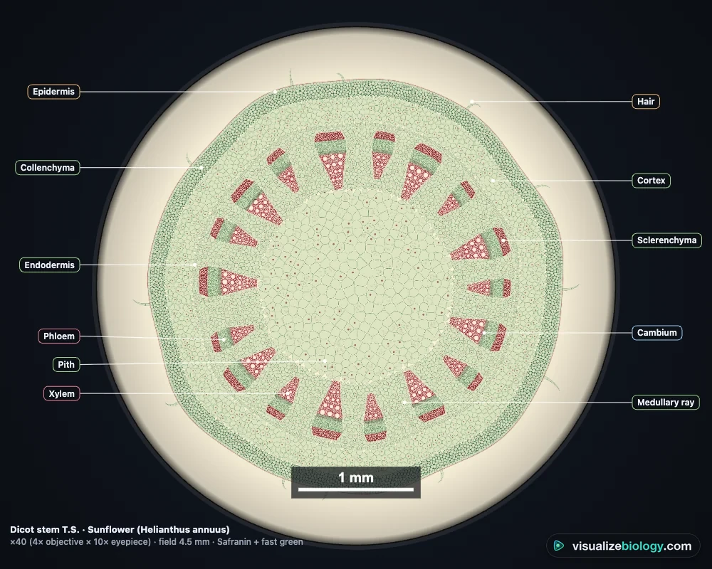
From the outside in, a young sunflower stem has an epidermis with a cuticle and hairs, a few layers of collenchyma, a cortex of parenchyma whose outer cells are green, and a starch sheath (the stem's endodermis). Then comes a ring of 15 to 20 vascular bundles separated by medullary rays. Each bundle is collateral and open: a cap of sclerenchyma fibres, phloem, a thin strip of cambium and xylem, with the narrow protoxylem toward the centre (endarch). The middle is a large pith.
Monocot vs dicot stem
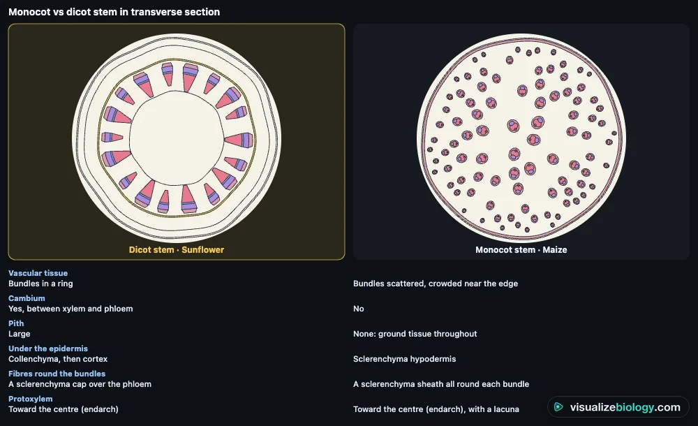
A maize stem looks completely different: under the epidermis is a hypodermis of sclerenchyma, and the rest is ground tissue with many small bundles scattered through it, crowded near the edge. Each bundle is closed (no cambium), wrapped in a sclerenchyma sheath, with two large metaxylem vessels and a protoxylem lacuna that make it look like a face.
| Feature | Dicot stem (sunflower) | Monocot stem (maize) |
|---|---|---|
| Vascular bundles | In a ring | Scattered, crowded near the edge |
| Kind of bundle | Collateral, open (cambium) | Collateral, closed (no cambium) |
| Under the epidermis | Collenchyma | Sclerenchyma hypodermis |
| Cortex, endodermis, pith | All present | Not distinct: ground tissue |
| Fibres | A cap over the phloem | A sheath round each bundle |
| Secondary growth | Yes | No |
Monocot vs dicot root
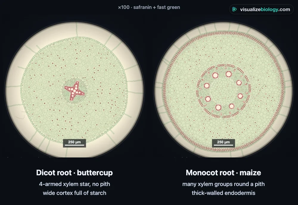
Both roots have a piliferous layer with root hairs, a wide cortex, an endodermis whose Casparian strips force water through cell membranes, and a pericycle where lateral roots begin. Inside, the xylem and phloem lie on different radii (radial bundles) with the protoxylem on the outside (exarch). A buttercup root has a four-armed xylem star (tetrarch) meeting in the centre, so it has no pith; a maize root has many xylem groups (polyarch) in a ring round a large pith.
How to tell a stem from a root
In a stem, each bundle has phloem outside xylem on the same radius and the protoxylem points inward (endarch). In a root the xylem and phloem alternate on different radii, the protoxylem points outward (exarch), and the endodermis is easy to see. Roots have no cuticle; young ones have root hairs.
How to draw a low-power plan
At ×40, draw only the boundaries of the tissues, never individual cells, in pencil with single continuous lines and in the right proportions. Label with ruled lines that do not cross, and give the magnification: size of the drawing ÷ actual size, in the same units. A 90 mm drawing of a 3.6 mm stem is ×25. The Plan view draws the plan for each section, with a scale bar.
Staining plant tissues
Phloroglucinol with hydrochloric acid turns lignin cherry red: xylem and sclerenchyma redden, collenchyma does not, because its thick walls are cellulose and pectin. Iodine turns starch blue-black, showing the starch sheath of a stem and the starchy cortex of a buttercup root. On prepared slides, safranin stains lignified walls red and fast green stains cellulose walls green.
Practicals and course links
| Practical | What you find out | Course links |
|---|---|---|
| Identify four mystery sections | Stem or root, monocot or dicot | IGCSE, GCSE, NCERT 11 |
| Draw and label a plan | Tissue positions; magnification | IGCSE, A-level, IB |
| Test for lignin and starch | Which walls are lignified | A-level, AP, college |
| Count the vascular bundles | Ring vs scattered; bundles per mm² | IGCSE, A-level, AP |
| Measure xylem vessels | Graticule calibration | A-level, IB |
| Root or stem? Count the xylem groups | Exarch; tetrarch and polyarch | A-level, NCERT, college |
Frequently asked questions
What is the difference between a monocot and a dicot stem?
In a dicot stem such as sunflower the vascular bundles form a ring, each open with cambium between its xylem and phloem, round a large pith, with collenchyma and a cortex outside them. In a monocot stem such as maize many closed bundles (no cambium) are scattered through ground tissue, crowded near the edge, each wrapped in a sclerenchyma sheath; there is a sclerenchyma hypodermis and no separate cortex or pith.
What is the difference between a monocot and a dicot root?
Both have radial bundles: xylem and phloem alternate on different radii, with the protoxylem on the outside (exarch), inside an endodermis and pericycle. A dicot root such as buttercup has few xylem arms (2 to 6; 4, tetrarch, in buttercup) that meet in the centre, so there is no pith. A monocot root such as maize has many xylem groups (polyarch, 12 to 20 or more) in a ring round a large pith.
What are the types of plant tissues?
Meristems make new cells; the permanent tissues are dermal (the epidermis), ground (parenchyma, collenchyma and sclerenchyma) and vascular (xylem and phloem). Parenchyma, collenchyma and sclerenchyma are simple tissues, made of one kind of cell; xylem and phloem are complex tissues, made of several kinds.
How do you tell a stem from a root in a cross section?
Look at the vascular tissue. In a stem each bundle has phloem outside xylem on the same radius (collateral) and the protoxylem points to the centre (endarch). In a root, xylem and phloem lie on different radii and alternate (radial), the protoxylem faces outward (exarch), and a clear endodermis with Casparian strips surrounds them; roots also have root hairs instead of a cuticle.
Explore related simulators
The microscope here is the engine of the Virtual Microscope, where you learn to focus, calibrate the graticule and look at onion, cheek and blood cells. To see what the xylem and phloem in these sections actually carry, follow water and sugar through a whole plant in Xylem and Phloem. The leaf's own tissues, its palisade, spongy mesophyll and stomata, are in the Leaf Structure & Stomata Virtual Lab, and how monocots and dicots fit into the plant kingdom is in the Plant Classification Game.
Sources: Evert, Esau's Plant Anatomy (3rd ed., Wiley 2006); Esau, Anatomy of Seed Plants (2nd ed., Wiley 1977); Raven, Evert and Eichhorn, Biology of Plants (8th ed.), chapters 23–26; NCERT Biology Class 11, chapter 6; OpenStax Biology 2e, chapter 30. An educational model: real sections vary with the plant, its age and the cut.
Updated