
Leaf Structure and Stomata Virtual Lab: Leaf Cross Section, Peel and Stomata Count
Look inside a bean, privet, maize, water lily or marram grass leaf in section, in 2D or 3D, with every cell drawn to scale. Watch the guard cells open the stomata at dawn and close them in a drought, make a nail varnish peel, count the stomata per mm², and run six leaf practicals.
What's happening
Lab notebook
Stomata opening and closing, midday closure, wilting, your counts and measurements and every condition you change are written here with the time. Export a CSV, or turn the session into a PDF lab report.
| # | Leaf | What | Result | Value |
|---|
Concept library
Click "New problem" to begin.
Click "Start quiz" for 5 questions on leaf structure, stomata, gas exchange or adaptations, and a calculation.
1 Overview
Simulate shows a leaf on the left, in 2D or 3D, and close-ups on the right: one Stoma face on, Gases diffusing through it, a Graph through the day and a dicot and monocot leaf to Compare. One model drives everything: the guard cells, the pores, water loss and photosynthesis.
2 Leaf, section and peel
- Leaf shows the whole leaf and the line the section is cut along (tap it to go to the section).
- Section is a transverse section with every cell drawn to scale in µm. Hold Ctrl or ⌘ and scroll to zoom, then drag to move. Tap any tissue to open its card on the right.
- Peel is the epidermis from above under the microscope. Choose the upper or lower surface and the ×100 or ×400 objective in the Leaf panel, and move the slide to see another field.
3 The day and the conditions
- Press ▶ (or Space) to run the clock. The strip under it shows night, dawn, morning, midday and evening: click one to jump there. ↺ starts a new day at 05:00.
- In Conditions set the light, CO₂, air humidity, wind, soil water and temperature, or spray ABA. The stomata take minutes to respond, as in a real leaf.
- Gas flow shows CO₂ coming in and water vapour going out at the model's rates.
4 Experiment mode and the lab report
Six practicals: label a leaf section, a nail varnish peel with stomata counts on both surfaces, the stomatal index of sun and shade leaves, measuring a stoma with an eyepiece graticule, epidermal strips on solutions, and leaves coated with petroleum jelly. Each shows its aim, what you need and a real-lab safety note; the method ticks off as you work, and a conclusion question unlocks the PDF lab report.
5 Units and keyboard
- SI | US switches between °C and °F (and millimetres and inches in the text); cells, pores and densities stay in µm and per mm², as under any microscope.
- Space plays or pauses, → jumps to the next part of the day, 1, 2 and 3 choose Leaf, Section or Peel, F shows or hides the gas flow, N starts a new day and S opens the live readings.
A leaf structure and stomata virtual lab in your browser
This virtual lab shows a leaf the way you see it under a microscope, but alive. Choose a bean, privet, maize, water lily or marram grass leaf and look at it in transverse section, in 2D or 3D, with every cell drawn to scale in micrometres. Then look at its surface from above, as a nail varnish peel, and watch the stomata open at dawn and close in the evening or in a drought. Behind it is a model of the guard cells as osmotic valves, built from plant physiology data, so light, CO₂, humidity, wind, soil water and the hormone ABA change the pores, the water lost and the sugar made.
- Leaves
- Bean, privet, maize, water lily and marram grass: dicot, monocot, hydrophyte and xerophyte, sun or shade
- Views
- Whole leaf, a cross section to scale, the epidermis at ×100 and ×400, one stoma close up
- Conditions
- Light (and its color), CO₂, humidity, wind, soil water, temperature and ABA
- Modes
- Simulate, Experiment (6 practicals with a PDF report) and Quiz
- Price
- Free, with no account or download; works on Chromebooks, tablets and phones
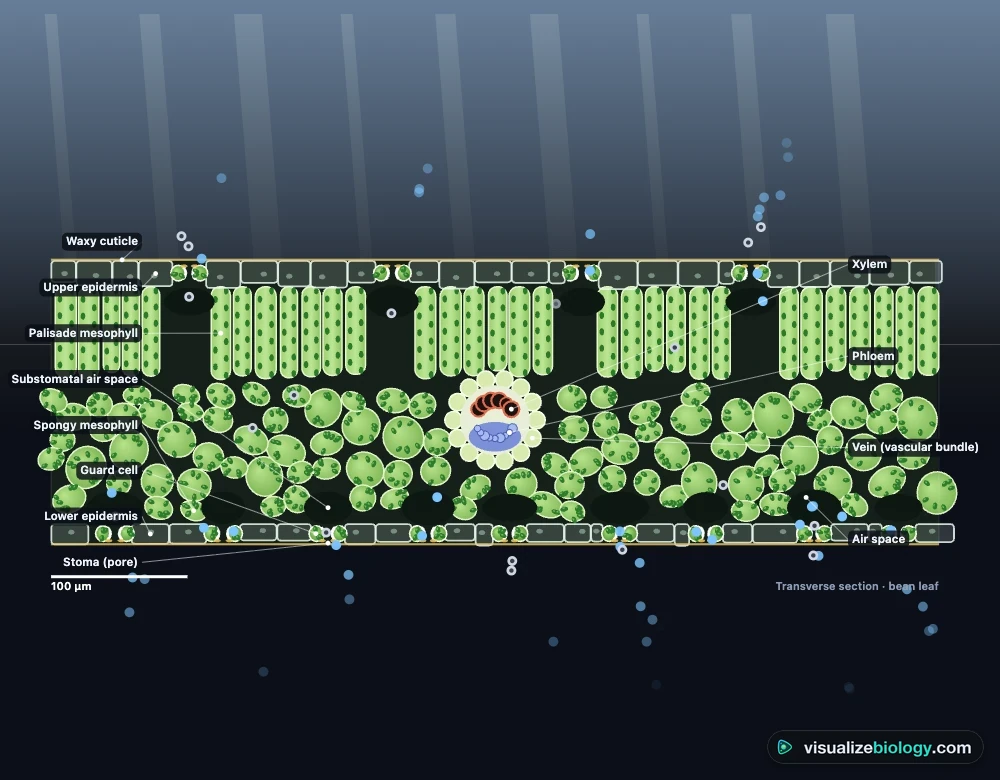
Leaf cross section labeled: the layers of a leaf
| Layer | What it looks like | Its job |
|---|---|---|
| Waxy cuticle | A clear, waxy film on top of the epidermis | Cuts water loss by evaporation |
| Upper epidermis | One layer of flat cells with no chloroplasts | Protects the leaf and lets light through |
| Palisade mesophyll | Tall, packed cells full of chloroplasts | Most of the photosynthesis |
| Spongy mesophyll | Rounded, loose cells with air spaces | Gas exchange: CO₂ reaches every cell |
| Vein (vascular bundle) | Xylem above, phloem below, in a sheath | Water in, sugar out, support |
| Lower epidermis | Epidermal cells with many stomata | Gas exchange through the stomata |
| Guard cells and stoma | Two bean-shaped cells around a pore | Open and close the pore |
How guard cells open and close stomata
Each stoma is a pore between two guard cells. In light, especially blue light, proton pumps push H⁺ out of the guard cells and potassium ions flow in, with chloride and malate. The water potential inside falls, water enters by osmosis and the guard cells become turgid. Their inner walls are thicker and cellulose fibres wrap around them like hoops, so they bow apart and the pore opens. In the dark, at high CO₂, in dry air or when the leaf is short of water (through the hormone ABA), potassium leaves, water follows and the pore closes. Open stomata let CO₂ in for photosynthesis but let water vapour out: every leaf trades water for sugar.
How to count stomata: the nail varnish peel
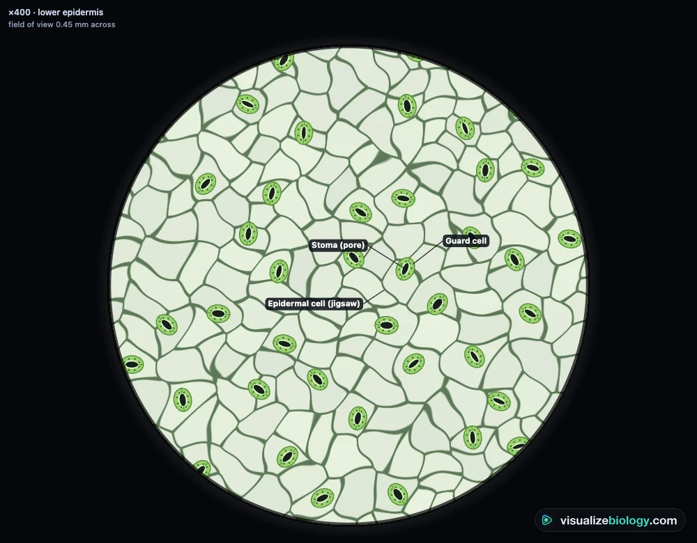
Paint a thin patch of clear nail varnish on the lower surface of a leaf, avoiding the midrib, and let it dry. Press clear sticky tape onto it, peel it off and stick it to a slide: the varnish is a cast of the surface. At ×400 count the stomata in the field of view. With a ×10 eyepiece and ×40 objective the field is about 0.45 mm across, so its area is π × 0.225² ≈ 0.159 mm². Divide the count by that area for the stomatal density, and repeat in several fields. The stomatal index is stomata ÷ (stomata + other epidermal cells) × 100; it does not change when cells grow bigger, which is why A-level practicals compare it between leaves.
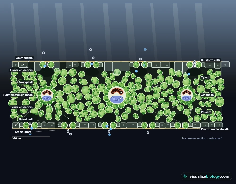
Monocot vs dicot leaf
| Dicot (bean, privet) | Monocot (maize, grasses) | |
|---|---|---|
| Veins | A branching net | Parallel |
| Mesophyll | Palisade above, spongy below | All alike, no palisade |
| Stomata | Mostly on the lower surface | Often on both surfaces, in rows |
| Guard cells | Bean shaped | Dumbbell shaped, with subsidiary cells |
Leaves adapted to dry and wet places
Marram grass grows on sand dunes: a thick cuticle, a leaf that rolls up in dry air, stomata sunk in grooves between hairs, and hinge cells that do the rolling. All of these trap humid air around the stomata, so less water diffuses out. A water lily floats: its stomata are only on the upper surface and huge air spaces (aerenchyma) keep it afloat. Maize, a C4 plant, gets more sugar per drop of water than a bean.
Practicals and course links
| Practical | What you find out | Course links |
|---|---|---|
| Label a leaf cross section | The tissues of a dicot and a monocot leaf | AQA GCSE 4.2.3.1; IGCSE 6.2 |
| Nail varnish peel | Stomata per mm² on each surface | GCSE; A-level stomatal count |
| Sun and shade leaves | Stomatal density and index | A-level; AP Bio |
| Eyepiece graticule | Calibration and the size of a stoma | GCSE and A-level microscopy |
| What opens stomata? | Light, K⁺, ABA and osmosis | A-level; AP Bio 8.1; IB |
| Petroleum jelly leaves | Which surface loses water | GCSE; IGCSE 7 |
Frequently asked questions
What are the layers of a leaf cross section?
From top to bottom: a waxy cuticle, the upper epidermis (transparent cells with no chloroplasts), the palisade mesophyll (tall cells packed with chloroplasts, the main site of photosynthesis), the spongy mesophyll (loose cells with air spaces for gas exchange), veins with xylem above and phloem below, and the lower epidermis with most of the stomata, each between two guard cells. Monocot leaves such as maize have no separate palisade and spongy layers.
How do guard cells open and close stomata?
In light, proton pumps in the guard cell membranes push H⁺ out, and potassium ions (with chloride) flow in. This lowers the water potential of the guard cells, water enters by osmosis and they become turgid. Their inner walls are thicker and cellulose fibres run around them, so they bow apart and the pore opens. In the dark, in a drought (through the hormone ABA) or at high CO₂, potassium leaves, water follows and the stomata close.
How do you calculate stomatal density from a nail varnish peel?
Count the stomata in the field of view, then divide by the area of the field: area = π × r². With a ×10 eyepiece and a ×40 objective the field is about 0.45 mm across, an area of about 0.159 mm², so 35 stomata means about 220 stomata per mm². Count several fields and take the mean. The stomatal index is stomata / (stomata + other epidermal cells) x 100.
Explore related simulators
What the palisade cells do with that light and CO₂: test light, CO₂ and temperature in the Photosynthesis Lab. The water lost through the stomata pulls water up the stem: follow it in the Xylem and Phloem Simulator. Guard cells open and close by osmosis: see water potential at work in the Osmosis Lab, and focus on real slides in the Virtual Microscope.
Sources: Franks and Beerling (2009), PNAS 106, for conductance from stomatal size and density; Roelfsema and Hedrich (2005), New Phytologist 167, for guard-cell signalling; Jarvis (1976), Phil. Trans. R. Soc. B 273, for the responses of stomata to light, CO₂ and humidity; Taiz, Zeiger et al., Plant Physiology and Development, 6th edition; OpenStax Biology 2e, chapter 30; Campbell Biology, 12th edition, chapters 35 and 36. This is an educational model: real stomata vary from place to place on a leaf.
Updated