
Virtual Stethoscope Trainer: Heart and Lung Sounds
Put the stethoscope on a virtual patient and listen: S1 and S2, splitting, gallops, murmurs, crackles and wheezes. Every sound is made by a working model of the heart and lungs, so it changes with where you listen, bell or diaphragm, and how the patient breathes.
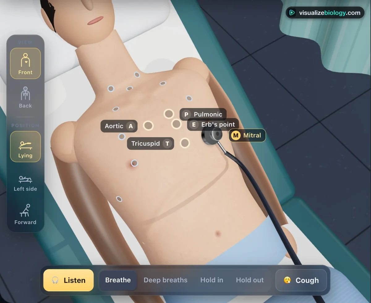
Listening posts
1 Overview
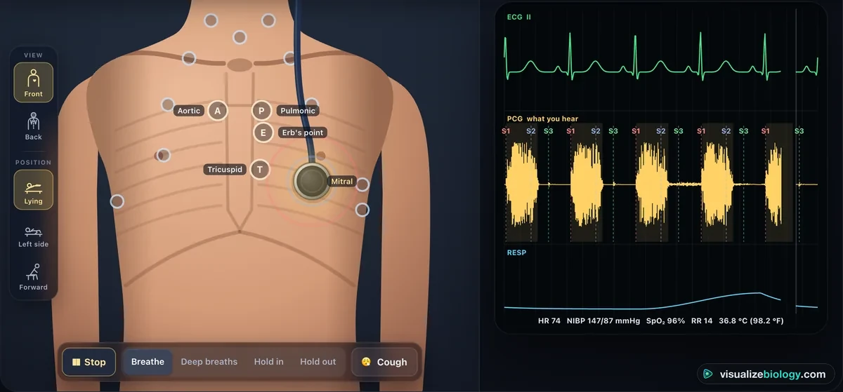
A virtual patient lies on the couch; the right-hand screen is the bedside monitor. Press 🎧 Listen, then move the chest piece: what you hear is computed, sample by sample, from a model of this patient's heart, circulation and lungs, heard at that spot through the stethoscope you chose. The monitor's sound trace (PCG) is the very sound you are hearing, lined up with the ECG. It is an educational model, not medical advice.
2 Listening
- Choose a patient in the command bar.
- Press 🎧 Listen (Space). Use headphones.
- Drag the chest piece, click a listening post, or use the Listening posts buttons. Front | Back turns the patient.
- Ask the patient to breathe normally, take deep breaths, hold the breath in or out, or cough. Roll them onto the left side or have them lean forward.
3 The stethoscope
Six generic types: the dual-head classic most students buy, a tunable single head, a dual-lumen cardiology scope, a pediatric scope, an electronic scope with filter modes, and a Sprague–Rappaport. The diaphragm turns the low sounds down; the bell, held lightly, passes them; press the bell hard and it acts like a diaphragm. A loose earpiece loses the lows and lets the room in. The Stethoscope tab of the monitor draws the response of what you are holding.
4 The monitor
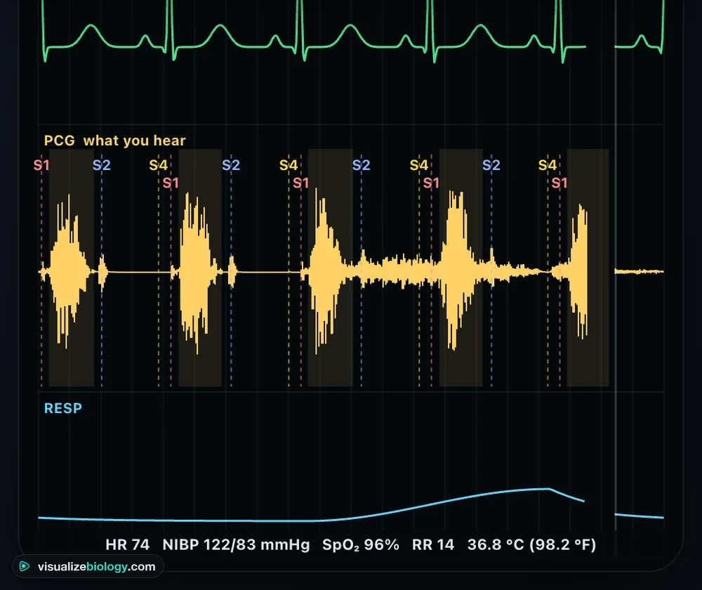
Monitor: ECG lead II, the sound trace (PCG) and the breathing, with heart rate, blood pressure, oxygen saturation, breathing rate and temperature. With "Name the sounds" on, S1, S2, S3, S4 and the opening snap are named where they happen, and a murmur is shaded. Sound: a spectrogram, low pitches at the bottom. Stethoscope: the frequency response.
5 Keys
- Space listen on/off · arrows move the chest piece 1 cm (Shift: 3 cm) · 1–5 aortic, pulmonic, Erb's, tricuspid, mitral · F / B front or back · D diaphragm or bell · N breathe normally · I deep breaths · H hold the breath · C cough.
How to use a stethoscope: heart and lung auscultation, step by step
Auscultation is listening to the body through a stethoscope. Heart sounds are made by valves closing and by blood speeding up and slowing down; murmurs by blood forced through a narrow or leaking valve; breath sounds by air rushing through the airways, filtered by the lung on its way to the chest wall. This free virtual stethoscope simulator makes all of them from a working model of the heart and lungs, so you hear each one change with where you listen, bell or diaphragm, how hard you press and how the patient breathes, the way it does at a real bedside.
It is built for students who need to recognise heart and lung sounds before they meet a patient: nursing students preparing for skills checks and the NCLEX, medical and physician assistant students, paramedics, and AP Biology or health science classes learning how the heart and lungs work. It runs in the browser on a laptop, Chromebook, tablet or phone; headphones help, because heart sounds sit low, between 20 and 200 Hz.
- Listen
- Drag the chest piece anywhere on the front or back, or jump to the 5 heart posts and 19 lung, neck and axilla sites
- Sounds
- S1, split S2, S3, S4, opening snap, 5 murmurs, vesicular, bronchial and tracheal breathing, fine and coarse crackles, wheezes
- Patients
- 16, from a healthy adult, athlete and child to heart failure, atrial fibrillation, pneumonia, asthma and lung fibrosis
- Stethoscopes
- 6 generic types: dual-head, tunable, cardiology, pediatric, electronic and Sprague–Rappaport
- Views
- 2D patient on a couch or a 3D examination room; monitor with ECG, sound trace and vitals; spectrogram
Heart auscultation points: the five listening posts
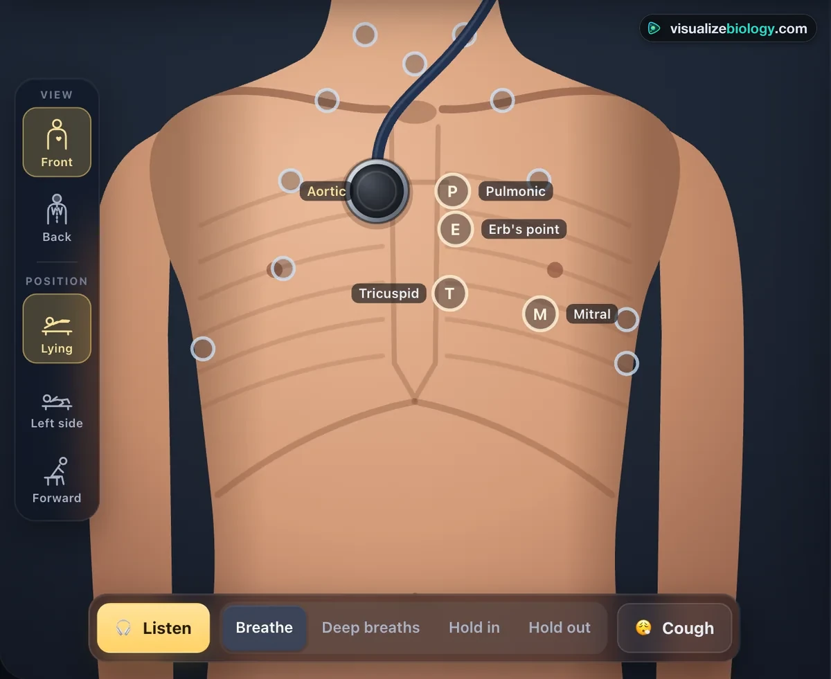
Each valve is heard best not over itself but where its sound and blood flow travel to the chest wall. Listen at the five posts in order, first with the diaphragm and then with the bell, with the patient lying back at about 30–45°:
| Post | Where on the chest | What you hear best |
|---|---|---|
| Aortic area | Right 2nd intercostal space at the sternal border | S2 (A2), the murmur of aortic stenosis, carried up to the neck |
| Pulmonic area | Left 2nd intercostal space at the sternal border | The split of S2 (A2 then P2) on breathing in |
| Erb's point | Left 3rd intercostal space at the sternal border | Aortic regurgitation, best with the patient sitting forward |
| Tricuspid area | Left 4th–5th intercostal space at the lower sternal border | S1, right-sided murmurs and gallops |
| Mitral area (apex) | Left 5th intercostal space in the midclavicular line | S1, S3 and S4, mitral regurgitation and the rumble of mitral stenosis |
Bell vs diaphragm: which side of the stethoscope?
Press the diaphragm (the large flat side) firmly for high-pitched sounds: S1, S2 and its split, most murmurs, breath sounds and crackles. Rest the bell (the small cup) lightly for low-pitched sounds: an S3 or S4 gallop and the rumble of mitral stenosis, best at the apex with the patient rolled onto the left side. Press the bell hard and the stretched skin turns it into a diaphragm, so the low sounds vanish; try it with the pressure slider. Always listen on bare skin: clothing rubbing on the chest piece sounds like crackles.
Normal heart sounds: S1, S2 and the split S2
S1 ("lub") is the mitral and tricuspid valves closing as the ventricles start to squeeze; S2 ("dub") is the aortic (A2) and pulmonary (P2) valves closing as they relax. Between S1 and S2 is systole, the short pause; after S2 comes diastole, the longer one. Breathing in pulls more blood into the right heart, so the pulmonary valve shuts a little later and S2 splits into two clicks a few hundredths of a second apart, which join again on breathing out. Ask the patient for deep breaths at the pulmonic area and watch the split come and go with the breathing trace.
| Sound | When | What makes it | Hear it |
|---|---|---|---|
| S1 | Start of systole, with the carotid pulse | Mitral and tricuspid valves closing | Apex, diaphragm |
| S2 | End of systole | Aortic (A2) and pulmonary (P2) valves closing | Base, diaphragm |
| Split S2 | Breathing in | P2 delayed by extra filling of the right heart | Pulmonic area, diaphragm |
| S3 | Early diastole, just after S2 ("Ken-tuck-y") | Blood rushing into a large or failing ventricle; normal in children and athletes | Apex, bell, patient on the left side |
| S4 | Late diastole, just before S1 ("Ten-nes-see") | The atrium pushing blood into a stiff ventricle, as in long-standing high blood pressure | Apex, bell |
| Opening snap | Soon after S2 | A stiff mitral valve snapping open in mitral stenosis | Apex and lower left sternal border |
Heart murmurs: what each one sounds like
A murmur is the rushing noise of blood forced through a narrow or leaking valve. Describe it by its timing (systolic or diastolic), shape, where it is loudest, where it spreads, its pitch and its grade from 1 (only just heard) to 6 (heard with the stethoscope lifted off the chest). In the trainer each murmur's pitch and loudness come from the flow and the pressure drop across the valve, so it grows and fades through the beat as a real one does.
| Murmur | Timing and shape | Loudest at | Clue |
|---|---|---|---|
| Aortic stenosis | Systolic, crescendo–decrescendo (diamond) | Aortic area, spreads to the neck | Harsh; S2 soft; an S4 is common |
| Mitral regurgitation | Holosystolic, flat, from S1 to S2 | Apex, spreads to the left armpit | Blowing; often an S3 |
| Aortic regurgitation | Early diastolic, decrescendo | Erb's point, patient sitting forward, breath held out | Soft and high; easy to miss |
| Mitral stenosis | Mid-diastolic rumble after an opening snap | Apex, bell, patient on the left side | Low; loud S1 |
| Still's (innocent) murmur | Early to mid systolic, musical | Lower left sternal border in a child | Vibratory, like a twanging string; the heart is normal |
How to listen to the lungs: auscultation landmarks
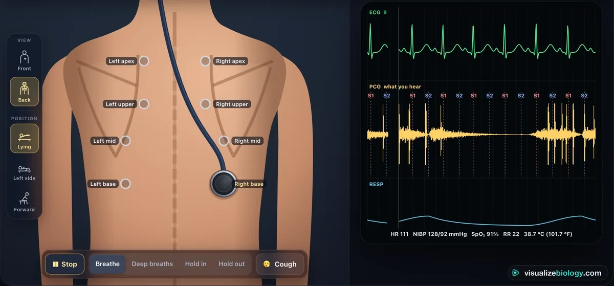
Ask the patient to breathe slowly and a little deeper than usual through an open mouth. Use the diaphragm and listen to at least one full breath at each site, moving side to side like a ladder so you always compare left with right at the same level. On the front, start above the clavicles at the apices, then the 2nd intercostal spaces, then the 4th on the right for the middle lobe, and the sides in the midaxillary line. Most of the lower lobes lie behind, so then listen to the back: have the patient cross their arms to move the scapulae aside, and go from the apices between the spine and the scapulae down to the bases, below the tips of the scapulae.
Normal lung sounds: vesicular, bronchovesicular and bronchial
Over most of the chest you hear soft, low vesicular breath sounds: breathing in is heard all the way, breathing out fades early, because the air-filled lung filters out the high pitches. Over the main bronchi, near the sternum and between the scapulae, the sound is bronchovesicular, with in and out about equal. Over the trachea it is tracheal: loud, harsh and hollow. Bronchial breathing heard over the lung is abnormal: when pneumonia fills the air spaces with fluid, the solid lung carries the airway's sound straight to the chest wall.
Abnormal lung sounds: crackles, wheezes, rhonchi and stridor
| Sound | What it sounds like | Cause | In the trainer |
|---|---|---|---|
| Fine crackles (rales) | Short, soft pops late in each breath in, like Velcro pulled apart | Small airways snapping open: lung fibrosis, fluid in heart failure | Pulmonary fibrosis, heart failure |
| Coarse crackles | Louder, lower bubbling early in the breath; may change after a cough | Mucus or fluid in larger airways: pneumonia, bronchiectasis | Pneumonia (try the cough button) |
| Wheezes | Musical, whistling notes, mostly breathing out; several at once in asthma | Air squeezing through narrowed airways | Asthma attack |
| Rhonchi | Low, snoring or rattling continuous sounds that often clear with a cough | Secretions in the large airways | Not in this version |
| Stridor | Loud, high, harsh sound on breathing in, heard best over the neck | A blocked upper airway: croup, a foreign body; an emergency | Not in this version |
| Pleural friction rub | Creaking like leather, on breathing in and out, in one spot | Inflamed pleura rubbing together: pleurisy | Not in this version |
The 3D examination room
Press 3D above the patient to move into a clinic room: an examination couch whose backrest rises and falls as you change the posture, an exam lamp, a curtain and a wall monitor with the live ECG and vitals. Drag the chest piece over the skin, walk round the couch to the back, and turn on the heart and lungs outline to see the organs under the listening posts. Lying, rolled onto the left side and sitting forward bring out the sounds that each posture is used for.
How to practise with the simulator
- Pick a patient and read why they have come in, then press Listen (or Space) and put on headphones.
- Go round the five heart posts with the diaphragm, then the apex again with the bell. Name S1 and S2 by their timing against the ECG, not by loudness.
- Ask for deep breaths, a held breath or a cough, and change the posture: listen for what changes.
- Turn the patient and work down the back from the apices to the bases, comparing left with right.
- Press "What should I hear?" for the teaching notes, and switch on "Name the sounds" to check yourself on the trace.
Frequently asked questions
When should you use the bell and when the diaphragm of a stethoscope?
Use the diaphragm, pressed firmly, for high-pitched sounds: S1 and S2, the splitting of S2, most murmurs (aortic stenosis, mitral and aortic regurgitation) and breath sounds and crackles. Use the bell, resting lightly on the skin, for low-pitched sounds: an S3 or S4 gallop and the rumble of mitral stenosis. Press the bell hard and the stretched skin turns it into a diaphragm, so the low sounds disappear.
Where are the five auscultation points of the heart?
The aortic area is the right 2nd intercostal space at the sternal border; the pulmonic area is the left 2nd intercostal space at the sternal border; Erb's point is the left 3rd intercostal space; the tricuspid area is the left 4th–5th intercostal space at the lower sternal border; and the mitral area (the apex) is the left 5th intercostal space in the midclavicular line.
How do you tell S1 from S2?
S1 (lub) comes just as the carotid pulse rises and is loudest at the apex; S2 (dub) is shorter, higher-pitched and loudest at the base. At normal heart rates the pause after S2 (diastole) is longer than the pause after S1 (systole).
What is the difference between crackles and wheezes?
Crackles (rales) are short, discontinuous pops, mostly while breathing in, made as closed small airways snap open or air bubbles through fluid: fine crackles in lung fibrosis and heart failure, coarse ones in pneumonia. Wheezes are continuous musical notes, mostly while breathing out, made by air squeezing through narrowed airways, as in asthma.
Where do you listen to the lungs with a stethoscope?
Listen at matching points on both sides, comparing left with right: on the front above the clavicles, at the 2nd and 4th intercostal spaces and at the sides in the midaxillary line; on the back from the apices down between the spine and the scapulae to the bases, below the tips of the scapulae. The lower lobes are heard mostly from the back.
Are the sounds in this stethoscope simulator real recordings?
No. Every sound is synthesised by an educational model of the heart, circulation and lungs, so it changes with the listening post, the chest piece, pressure, breathing and posture. It trains recognition of the sound patterns; it is not a recording of a patient or a diagnostic reference.
Explore related simulators
See the valves that make these sounds open and close in the Heart Simulator, take a blood pressure by ear in the Blood Pressure Simulator, and watch the lungs that make the breath sounds in the Respiratory System Simulator.
Sources: Bickley, Bates' Guide to Physical Examination; Tavel, Clinical Phonocardiography; Xu, Durand & Pibarot 2001 (the S2 model); Pasterkamp et al. 2018, ERS lung sound nomenclature; Sovijärvi et al. 2000 (CORSA); Abella et al. 1992 (stethoscope acoustics); Klabunde, Cardiovascular Physiology Concepts. Every sound here is synthesised by an educational model: it trains recognition and is not a recording of a patient or a diagnostic reference.
Updated