
Interactive 3D Teeth Chart: Names, Numbers, Types and Parts of a Tooth
Every adult and baby tooth from real scans, in the jaws in 3D or on a dental chart. Click a tooth to hold it on its own, turn it and cut it open, and read its name, its Universal, FDI and Palmer number and its job; then watch teeth come in and fall out from birth to adulthood.
This mouth
Lab notebook
The teeth you look at, the ages you explore and your practical results are written here. Export a CSV, or turn the session into a PDF lab report.
| # | When | What | Result | Value |
|---|
Concept library
Click "New problem" to begin.
Click "Start quiz" for 5 questions: types of teeth, tooth numbers, the parts of a tooth, baby and adult teeth, and decay.
1 Overview
Simulate shows the teeth on the left, as a 2D dental chart or front and side views, or in 3D in a real skull. The right card shows the tooth you pick: Tooth (that tooth alone in 3D with its details), Inside (a real section with its layers), Ages (when every tooth comes in and falls out) and Decay (sugar, plaque acid and a cavity over time).
2 Picking and finding a tooth
- Click any tooth or its number badge, in 2D or 3D. The right card shows it alone: drag to turn it, use Front, Side and Top, switch on Labels (off at first) to name its parts and, separately, Surfaces to name its mesial, distal, facial and lingual surfaces, and switch on Cut open to see its pulp and canals. Click any part named on the tooth or in its parts list to read about it.
- Find a tooth lists every tooth as a button by quarter of the mouth; type a number (30, #19, 46, UR6, T) or a name (canine, wisdom) in the search box.
- Universal | FDI | Palmer in the command bar changes every number on the page.
3 Views, types and ages
- Chart, Front, Side, Upper and Lower choose the view (keys 1–5); in 3D, Iso (key 0) is the three-quarter view the page opens with. The rail on the left of the teeth shows one Type (All · Inc · Can · PM · Mol) and, in 3D, makes the Jaw bone see-through, solid or hidden and Opens the mouth (+ / −).
- Colour by type colours incisors, canines, premolars and molars; Type on the rail shows one type only.
- Adult · 32, Baby · 20 or By age: drag the age slider or press ▶ to watch teeth form in the jaw, come in and fall out from birth to 25.
4 Decay
Set sugary snacks a day, brushing, fluoride toothpaste and a fissure sealant, then run up to three years. The Decay card shows plaque pH through one day (the Stephan curve) and the depth of a cavity in the picked tooth through enamel, dentin and pulp; the tooth itself darkens in 2D, in 3D and in its section. An educational model, not dental advice.
5 Experiment mode and the lab report
Six practicals: name the teeth, count teeth and the dental formula, a child's dental age, eggshells in drinks, sugar and plaque pH, and a plaque score with a disclosing tablet. Each shows its aim, what you need and a safety note; the method ticks off as you work, and a conclusion question unlocks the PDF lab report.
6 Units and keyboard
- SI | US switches tooth sizes between mm and inches, bite force between newtons and pounds-force and eggshell mass between grams and ounces. Ages, pH and minutes are the same in both.
- Space plays the years, 0–5 choose the view (0 = the opening three-quarter view, in 3D), F jumps to Find a tooth, ←/→ step to the neighbouring tooth, C cuts the tooth open, S opens the mouth at a glance.
An interactive teeth chart built from real teeth
A teeth chart names and numbers every tooth in the mouth. This one is interactive and three-dimensional: each of the 32 adult and 20 baby teeth is a real tooth model, sculpted from CT scans by the University of Dundee School of Dentistry, set in the jaws of a real skull. Click a tooth and it appears on its own beside the chart: turn it to see its cusps and roots, cut it open to its pulp, and read its name, its number in all three numbering systems, what it does and when it comes in.
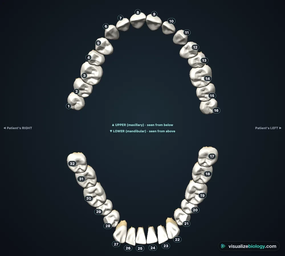
- Teeth
- 32 adult and 20 baby teeth from CT-based models (University of Dundee, CC BY 4.0) in a BodyParts3D skull
- Numbering
- Universal (1–32, A–T), FDI / ISO 3950 (11–48, 51–85) and Palmer
- Per tooth
- Name, type, job, crown and root length, roots, cusps, canals, bite force, eruption age, a 3D view and a real section
- Courses
- KS2 Year 4 teeth, IGCSE 7.3, NGSS MS-LS1-3, NCERT Class 7 and 11, dental assisting
- Price
- Free, with no account or download; works on Chromebooks, tablets and phones
The four types of teeth and their functions
Humans are heterodont: our teeth have different shapes for different jobs. Incisors, the eight flat front teeth, cut and bite like chisels. The four canines are pointed, with the longest roots in the mouth (the upper canine is about 27 mm long), to grip and tear. The eight premolars have two cusps and crush and tear; baby teeth have none. The twelve molars, including the four wisdom teeth, have broad chewing surfaces with four or five cusps that grind food, and they take the biggest bite forces, about 600 N against about 150 N for an incisor. Switch on Colour by type to see the four groups at once.
Teeth numbers and names: Universal, FDI and Palmer
In the United States teeth are numbered with the Universal system: 1 is the upper right third molar, the numbers run along the upper teeth to 16, the upper left third molar, then drop to the lower left third molar (17) and run back to the lower right third molar (32). Baby teeth are lettered A to T in the same order. Most of the world, including Europe, uses the two-digit FDI system (ISO 3950): the first digit is the quarter of the mouth (1 upper right, 2 upper left, 3 lower left, 4 lower right; 5–8 for baby teeth), the second the tooth counted from the midline, so 46 is the lower right first molar. In the UK the Palmer notation writes the same tooth as 6 in a bracket that shows its quarter. Every tooth on the chart has all three.
| Universal | FDI | Palmer | Tooth | Type | Comes in |
|---|---|---|---|---|---|
| 1 | 18 | 8┘ | Upper right third molar | Molar | 17–21 y |
| 2 | 17 | 7┘ | Upper right second molar | Molar | 12–13 y |
| 3 | 16 | 6┘ | Upper right first molar | Molar | 6–7 y |
| 4 | 15 | 5┘ | Upper right second premolar | Premolar | 10–12 y |
| 5 | 14 | 4┘ | Upper right first premolar | Premolar | 10–11 y |
| 6 | 13 | 3┘ | Upper right canine | Canine | 11–12 y |
| 7 | 12 | 2┘ | Upper right lateral incisor | Incisor | 8–9 y |
| 8 | 11 | 1┘ | Upper right central incisor | Incisor | 7–8 y |
| 9 | 21 | └1 | Upper left central incisor | Incisor | 7–8 y |
| 10 | 22 | └2 | Upper left lateral incisor | Incisor | 8–9 y |
| 11 | 23 | └3 | Upper left canine | Canine | 11–12 y |
| 12 | 24 | └4 | Upper left first premolar | Premolar | 10–11 y |
| 13 | 25 | └5 | Upper left second premolar | Premolar | 10–12 y |
| 14 | 26 | └6 | Upper left first molar | Molar | 6–7 y |
| 15 | 27 | └7 | Upper left second molar | Molar | 12–13 y |
| 16 | 28 | └8 | Upper left third molar | Molar | 17–21 y |
| 17 | 38 | ┌8 | Lower left third molar | Molar | 17–21 y |
| 18 | 37 | ┌7 | Lower left second molar | Molar | 11–13 y |
| 19 | 36 | ┌6 | Lower left first molar | Molar | 6–7 y |
| 20 | 35 | ┌5 | Lower left second premolar | Premolar | 11–12 y |
| 21 | 34 | ┌4 | Lower left first premolar | Premolar | 10–12 y |
| 22 | 33 | ┌3 | Lower left canine | Canine | 9–10 y |
| 23 | 32 | ┌2 | Lower left lateral incisor | Incisor | 7–8 y |
| 24 | 31 | ┌1 | Lower left central incisor | Incisor | 6–7 y |
| 25 | 41 | 1┐ | Lower right central incisor | Incisor | 6–7 y |
| 26 | 42 | 2┐ | Lower right lateral incisor | Incisor | 7–8 y |
| 27 | 43 | 3┐ | Lower right canine | Canine | 9–10 y |
| 28 | 44 | 4┐ | Lower right first premolar | Premolar | 10–12 y |
| 29 | 45 | 5┐ | Lower right second premolar | Premolar | 11–12 y |
| 30 | 46 | 6┐ | Lower right first molar | Molar | 6–7 y |
| 31 | 47 | 7┐ | Lower right second molar | Molar | 11–13 y |
| 32 | 48 | 8┐ | Lower right third molar | Molar | 17–21 y |
Parts of a tooth
A tooth has a crown above the gum, a neck at the cemento-enamel junction and one to three roots held in the jaw. The crown is covered by enamel, the hardest substance in the body (96 % mineral), up to about 2.5 mm thick over the cusps. Under it, dentin makes up most of the tooth; its tiny tubules are why an exposed tooth feels cold and sweet. In the middle the soft pulp, with blood vessels and nerves, runs down the root canals to the apical foramen at each root tip. The root is covered by cementum, which anchors the fibres of the periodontal ligament to the alveolar bone of the socket, and the gum seals the bone off from the mouth.
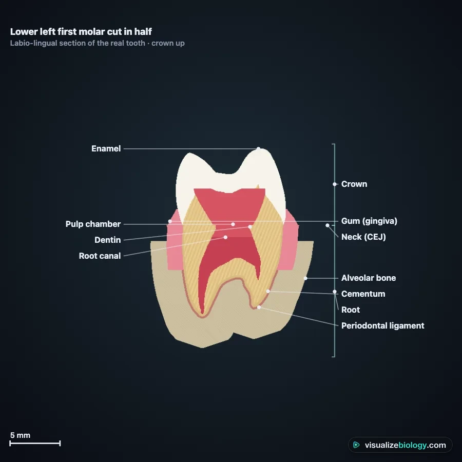
Baby teeth vs adult teeth: when they come in
Humans are diphyodont: we grow two sets of teeth. The 20 baby (primary) teeth come in from about 6 months, starting with the lower central incisors, and are all in by about 2½–3 years. From about 6 the first adult molars appear behind them, and the baby teeth are shed one by one as the adult teeth under them push up and dissolve their roots: this mixed dentition lasts until about 12. Baby molars are replaced by premolars, not molars. Use By age to watch it happen.
| Universal | FDI | Tooth | Comes in | Falls out |
|---|---|---|---|---|
| A | 55 | upper right second molar | 25–33 months | 10–12 y |
| B | 54 | upper right first molar | 13–19 months | 9–11 y |
| C | 53 | upper right canine | 16–22 months | 10–12 y |
| D | 52 | upper right lateral incisor | 9–13 months | 7–8 y |
| E | 51 | upper right central incisor | 8–12 months | 6–7 y |
| F | 61 | upper left central incisor | 8–12 months | 6–7 y |
| G | 62 | upper left lateral incisor | 9–13 months | 7–8 y |
| H | 63 | upper left canine | 16–22 months | 10–12 y |
| I | 64 | upper left first molar | 13–19 months | 9–11 y |
| J | 65 | upper left second molar | 25–33 months | 10–12 y |
| K | 75 | lower left second molar | 23–31 months | 10–12 y |
| L | 74 | lower left first molar | 14–18 months | 9–11 y |
| M | 73 | lower left canine | 17–23 months | 9–12 y |
| N | 72 | lower left lateral incisor | 10–16 months | 7–8 y |
| O | 71 | lower left central incisor | 6–10 months | 6–7 y |
| P | 81 | lower right central incisor | 6–10 months | 6–7 y |
| Q | 82 | lower right lateral incisor | 10–16 months | 7–8 y |
| R | 83 | lower right canine | 17–23 months | 9–12 y |
| S | 84 | lower right first molar | 14–18 months | 9–11 y |
| T | 85 | lower right second molar | 23–31 months | 10–12 y |
The human dental formula
A dental formula counts the teeth in one quarter of the mouth as incisors, canines, premolars and molars, upper over lower. For adults it is 2 1 2 3 / 2 1 2 3: 8 teeth per quarter × 4 = 32. For baby teeth it is 2 1 0 2 / 2 1 0 2 = 20. Our teeth also sit in sockets (thecodont). NCERT students: these are the three words in the Class 11 digestion chapter.
How tooth decay happens
Bacteria in plaque turn sugar into acid. After each sugary snack the plaque's pH falls within minutes and takes 30–60 minutes to recover (the Stephan curve). Below a critical pH of about 5.5, enamel loses mineral; above it, saliva puts mineral back, and fluoride makes that repair stronger and the enamel harder to dissolve. Decay is the running balance: frequent sugar wins, so a white spot becomes a cavity, spreads faster through the dentin and reaches the pulp. Brushing, fluoride and fewer sugary snacks tip the balance back. The eggshell practical shows the same chemistry: shell, like enamel, dissolves in acidic drinks. This is an educational model, not dental advice.
Wisdom teeth
The third molars come in last, usually between 17 and 21, if at all. About a quarter of people are missing at least one, and many jaws have no room, so a wisdom tooth stays impacted against the second molar, most often in the lower jaw. That is why many adults have 28 teeth rather than 32.
Practicals and course links
| Practical | What you find out | Course links |
|---|---|---|
| Name that tooth | Tooth names and numbers on a chart | KS2 Year 4, IGCSE 7.3, dental assisting |
| Count the teeth | Baby and adult dental formulas | NCERT Class 11, KS2 |
| How old is this child? | Dental age from erupted teeth | Forensic science, health science |
| Eggshells in drinks | Acid attack on a model of enamel | KS2 Year 4, NCERT Class 7 |
| Sugar and plaque pH | The Stephan curve and snack frequency | Health, GCSE chemistry links |
| Disclosing tablet | Plaque score before and after brushing | KS2, health education |
Frequently asked questions
How many teeth do adults have?
Adults have 32 permanent teeth: in each half of each jaw 2 incisors, 1 canine, 2 premolars and 3 molars (the dental formula 2123). The last molars are the wisdom teeth, which come in between about 17 and 21 years old, if at all; many people have only 28 teeth in the mouth because their wisdom teeth are missing or stay in the jaw. Children have 20 baby teeth (2102: no premolars, two molars).
What is tooth number 30?
In the Universal numbering system used in the United States, tooth 30 is the lower right first molar, the large back tooth that comes in at about 6 years old. Universal numbers run 1 to 16 along the upper teeth from the upper right wisdom tooth to the upper left one, then 17 to 32 back along the lower teeth from the lower left wisdom tooth to the lower right one. In the FDI system used in Europe the same tooth is 46.
What are the four types of teeth and their functions?
Incisors (8 in adults) are thin, chisel-shaped front teeth that cut and bite food. Canines (4) are pointed and have the longest roots; they grip and tear. Premolars (8, none in baby teeth) have two cusps and crush and tear. Molars (12 including wisdom teeth) are broad back teeth with four or five cusps that grind food before it is swallowed.
Explore related simulators
Teeth are where digestion starts: follow a mouthful from the teeth onward in the Human Digestive System Simulator. The skull and jaws that hold the teeth are in the Interactive 3D Skeleton, and the muscles that close the jaw, the masseter and temporalis, are in the Interactive 3D Muscular System. The acid that dissolves enamel comes from bacteria; see them grow in the Virtual Bacteria Simulator.
Sources: Nelson, Wheeler's Dental Anatomy, Physiology and Occlusion, 11th ed. (crown and root sizes); American Dental Association eruption charts (MouthHealthy); ISO 3950:2016 (FDI notation); Stephan (1944), J Dent Res 23: 257–266; Featherstone (2008), Aust Dent J 53: 286–291; OpenStax Anatomy and Physiology 2e, chapter 23. 3D teeth: University of Dundee School of Dentistry tooth morphology models (Sketchfab), CC BY 4.0, scaled to Wheeler's averages, simplified, mirrored for the right side and set on the arch of their own full-dentition model. Skull and jaws: BodyParts3D, The Database Center for Life Science (DBCLS), CC BY 4.0. This is an educational model, not dental advice.
Updated