
Leaf Pigment Chromatography Virtual Lab: Separate Chlorophylls and Carotenoids
A leaf is green, but it holds at least four pigments. Grind a leaf, spot the extract on paper or a TLC plate, and watch the solvent pull carotene, xanthophylls and chlorophylls a and b apart. Measure the bands, work out their Rf values, compare sun, shade, autumn and red leaves, and run AQA required practical 7 with no flammable solvent in the room.
What's happening
Lab notebook
Every step (lowering the paper in, taking it out, marking the front) and every band you measure is written here with its distance and Rf. Export a CSV, or turn the session into a PDF lab report.
| # | Lane | What | Result | Value |
|---|
Concept library
Click "New problem" to begin.
Click "Start quiz" for 5 questions on the leaf pigments, the method and working with Rf values.
1 Overview
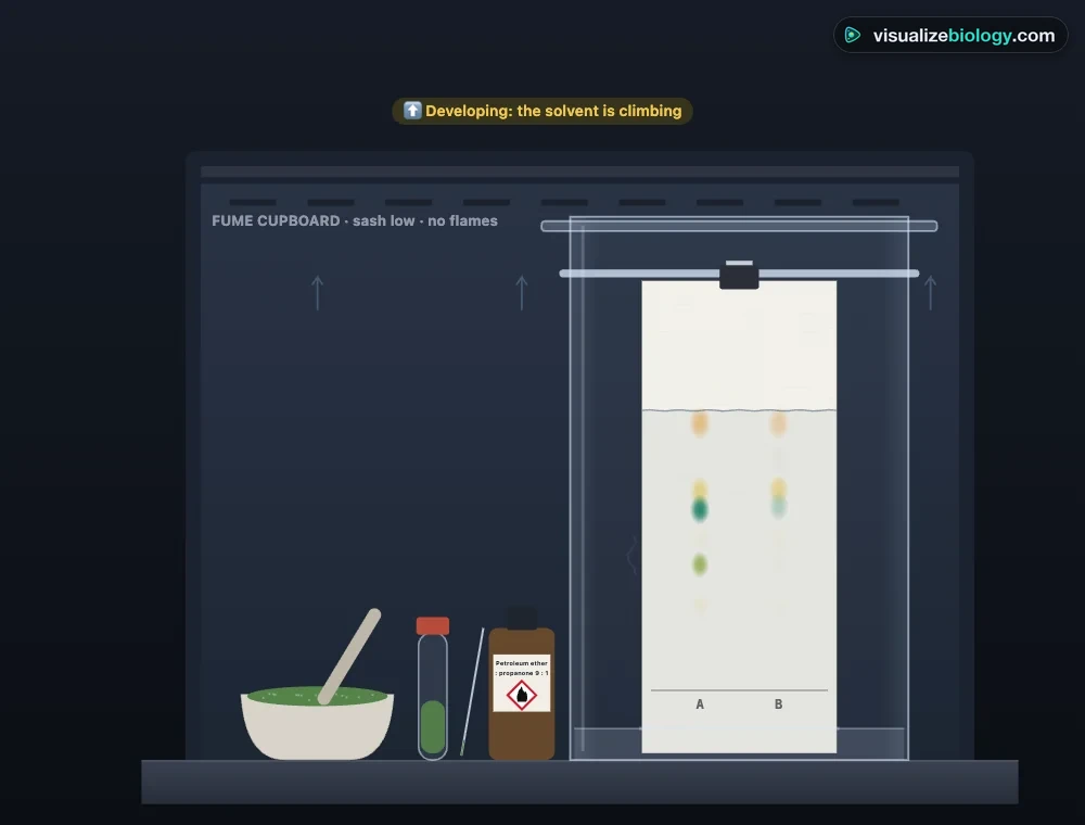
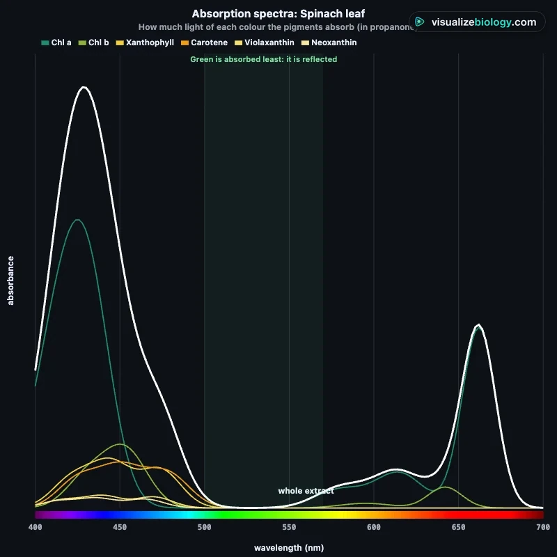
Simulate shows the bench on the left, inside a fume cupboard for the flammable solvents: a mortar of ground leaf, the extract, and a tank in which the paper develops. Bench and Paper choose the camera. On the right are close-ups: How it works (molecules sticking and dissolving), Pigments, Spectra and Graph. Tap any part or band to open its card.
2 Running a chromatogram
- Press ▶ (or Space). The paper is lowered into the solvent, which climbs it; the pigments separate into bands.
- With Do it for me (Method panel) the paper comes out about 1 cm from the top and the front is marked for you. With I'll do it, press Take out (O) and then Mark front (M) before the paper dries.
- In the Paper view, click the centre of each band to measure it. Its distance and Rf go into the notebook.
3 The set-up
- Leaves: up to four lanes (spinach, sun and shade ivy, autumn, red coleus, brown seaweed) and the number of drops on each spot.
- Method: the medium and solvent (also on the rail on the bench), five mistakes to switch on, and who takes the paper out.
4 Experiment mode and the lab report
Six practicals: spinach pigments (AQA required practical 7), sun and shade leaves, green, yellow and red leaves, choosing the solvent, paper or TLC, and four failed chromatograms. Each shows its aim, what you need and a real-lab safety note; the method ticks off as you work, and a conclusion question unlocks the PDF lab report.
5 Units and keyboard
- SI | US shows distances in mm or inches; Rf has no unit.
- Space plays or pauses, → skips to the next step, N starts a new run, O takes the paper out, M marks the front, 1 and 2 choose Bench and Paper, S opens the live readings.
A leaf pigment chromatography virtual lab
Chromatography of leaf pigments is one of the first separations most biology students do, and one of the most striking: a dark green spot turns into a ladder of orange, yellow, blue-green and yellow-green bands. This virtual lab runs the whole practical in your browser. Grind a leaf with sand and propanone, put the extract on a pencil origin line, and lower the paper into a shallow solvent in a closed tank. The solvent climbs by capillarity and carries each pigment its own distance, so they separate. You take the paper out, mark the solvent front, measure each band and work out its Rf value. A model of chromatography sits behind the drawing, so changing the leaf, the paper or the solvent changes the result as it would at the bench.
- Leaves
- Spinach, sun and shade ivy, a yellow autumn leaf, a red coleus leaf, a brown seaweed
- Media
- Chromatography paper, silica TLC plate, coffee filter; five solvents from water to petroleum ether
- Courses
- AQA A-level required practical 7; OCR, IB and AP Biology photosynthesis
- Modes
- Simulate, Experiment (6 practicals with a PDF report) and Quiz
- Price
- Free, with no account or download; works on Chromebooks, tablets and phones
How leaf pigment chromatography works
Chromatography separates a mixture between two phases. The stationary phase is the paper (cellulose holding a film of water) or the silica of a TLC plate; both are polar. The mobile phase is the solvent, usually petroleum ether with a little propanone, which is mostly non-polar. Each pigment molecule keeps sticking to the stationary phase and dissolving back into the moving solvent. The share of time it spends dissolved sets how far it travels. β-carotene has no oxygen atoms, so it barely sticks and runs almost with the front. Xanthophylls carry oxygen atoms and chlorophylls a polar head, so they lag behind. The How it works close-up shows this molecule by molecule.
Rf values of the leaf pigments
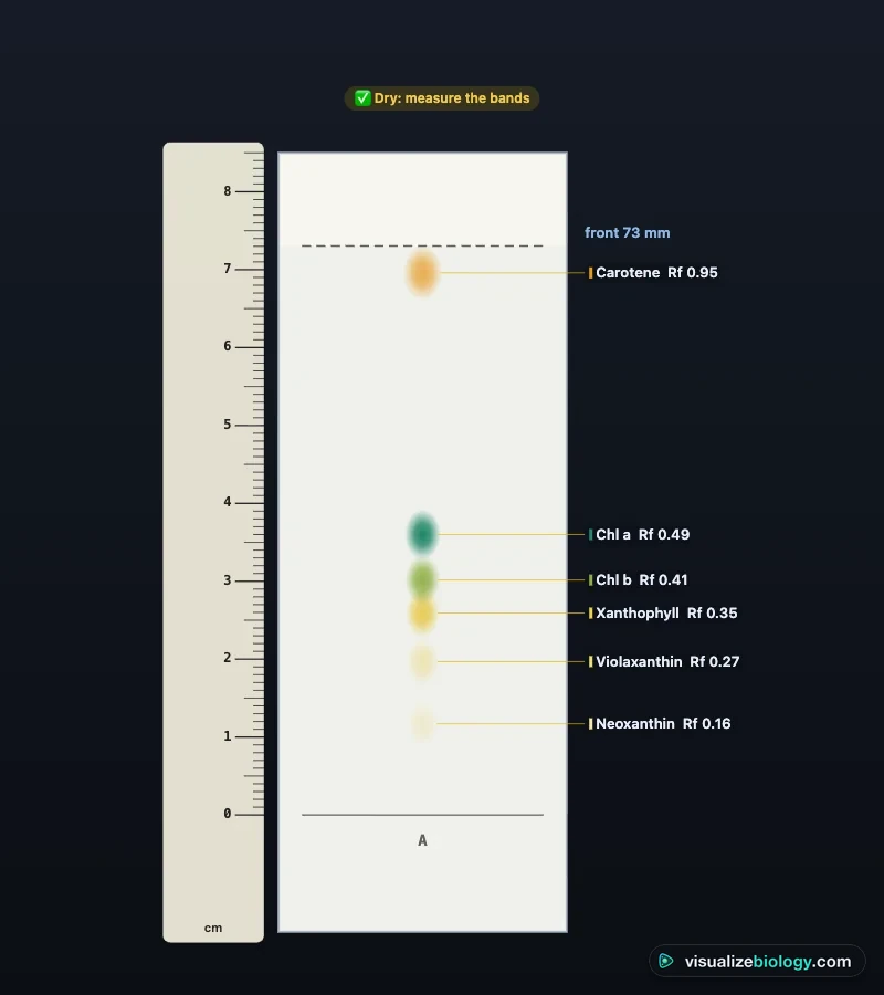
Rf = distance moved by the pigment ÷ distance moved by the solvent, both measured from the origin line to the centre of the band and to the front. It has no unit and lies between 0 and 1. The typical values below are for chromatography paper in petroleum ether : propanone (9 : 1). They change with the paper, the solvent and the temperature, so compare Rf values only within one system.
| Pigment | Colour | Rf on paper | Role |
|---|---|---|---|
| β-carotene | Yellow-orange | about 0.95 | Accessory pigment; protects against excess light |
| Xanthophyll (lutein) | Yellow | about 0.71 | Accessory pigment; sheds excess energy as heat |
| Chlorophyll a | Blue-green | about 0.65 | Primary pigment in the reaction centres |
| Chlorophyll b | Yellow-green | about 0.45 | Accessory pigment in the light-harvesting antenna |
Method: AQA required practical 7
Tear about 2 g of leaf and grind it with a little sand and 5 cm³ of propanone, which dissolves the pigments out of the thylakoid membranes. Draw a pencil line 2 cm from the bottom of the paper. With a capillary tube, put a small drop of extract on the line, dry it with cool air and repeat five to ten times, so the spot is concentrated but no wider than 3 mm. Hang the paper in a tank with about 1 cm of solvent, below the line, and close the lid. When the front is about 1 cm from the top, take the paper out and mark the front in pencil straight away: it dries and vanishes within a minute or two. Measure each band and the front from the origin, then calculate Rf.
Paper vs thin-layer chromatography (TLC)
A TLC plate carries a thin layer of fine, even silica gel. Its bands spread less than on coarse paper, so pigments with close Rf values separate: spinach gives four clear bands on paper but six on TLC, with violaxanthin and neoxanthin resolved. TLC is also faster. Silica holds the oxygen-rich xanthophylls more strongly than paper does, so on a plate they run below the chlorophylls.
Sun and shade leaves, autumn and red leaves
AQA suggests comparing leaves of different plants or colours. Shade leaves have relatively more chlorophyll b (a chlorophyll a : b ratio near 2.4, against about 3.5 in sun leaves). It sits in the light-harvesting antenna and catches more of the dim light that has passed through other leaves. A yellow autumn leaf has lost most of its chlorophyll, which reveals the carotenoids that were there all summer. A red leaf still has its chlorophyll: red anthocyanin, a water-soluble vacuole pigment, masks it and stays on the origin in a non-polar solvent.
Common mistakes and safety
Draw the origin in pencil: ink dyes dissolve and add false bands. Keep the solvent below the origin, or the spot dissolves into the tank. Keep the lid on, or the solvent evaporates from the paper and the front stalls and curves. A big spot gives wide, overlapping bands, and an extract left in light turns chlorophyll into grey pheophytin. Each of these can be switched on in the simulator. Petroleum ether, hexane and propanone are highly flammable and harmful, so in a real lab use them only in a fume cupboard, away from flames, with goggles and gloves.
Practicals and course links
| Practical | What you find out | Course links |
|---|---|---|
| Separate the pigments of spinach | Four pigments and their Rf values | AQA A-level RP7; IB C1.3; AP Bio 3.5 |
| Sun and shade leaves | Chlorophyll a : b ratio | AQA A-level RP7 |
| Green, yellow and red leaves | Masked carotenoids and anthocyanin | GCSE, AP Bio, A-level |
| Choosing the solvent | Polarity and solubility | A-level, IB |
| Paper or TLC? | Resolution of the bands | A-level, IB, college |
| What went wrong? | Diagnosing method errors | All practical exams |
Frequently asked questions
What are the Rf values of the leaf pigments?
On chromatography paper in petroleum ether : propanone (9 : 1), typical values are β-carotene about 0.95 (yellow-orange), xanthophyll about 0.71 (yellow), chlorophyll a about 0.65 (blue-green) and chlorophyll b about 0.45 (yellow-green). Rf values depend on the paper or plate, the solvent and the temperature, so they are only comparable within one system: on a silica TLC plate the xanthophylls run below the chlorophylls.
Why does carotene travel furthest in leaf chromatography?
β-carotene is a hydrocarbon with no oxygen atoms, so it is the least polar pigment. It dissolves best in the non-polar solvent and is attracted least to the polar paper or silica, so it spends almost all its time moving with the solvent. Xanthophylls carry oxygen atoms and chlorophylls have a polar head, so they are held back more and have lower Rf values.
Why is leaf pigment chromatography done in a fume cupboard?
The usual solvents, petroleum ether, hexane and propanone, are highly flammable and their vapour is harmful to breathe; the vapour is heavier than air and can travel to a flame. The tank is kept closed and used in a fume cupboard with no naked flames, with eye protection and gloves. A virtual lab lets you practise the method and the calculations without these hazards.
Explore related simulators
The pigments you separate here capture the light for photosynthesis: change light, CO₂ and temperature and follow the light reactions in the Photosynthesis Lab. The chloroplasts that hold them fill the palisade cells: cut a leaf in section and count its stomata in the Leaf Structure & Stomata Virtual Lab. Separating DNA fragments by size works on a similar idea of different speeds through a medium: run a gel in the Gel Electrophoresis Virtual Lab.
Sources: AQA A-level Biology required practical 7; Lichtenthaler (1987), Methods in Enzymology 148, 350–382; Sjursnes, Kvittingen and Schmid (2015), Journal of Chemical Education 92, 193–196; Taiz, Zeiger et al., Plant Physiology and Development, 6th ed., chapter 7; OpenStax Biology 2e, chapter 8. Rf values are typical teaching values; this is an educational model.
Updated