
Hematopoiesis Simulator: Watch the Bone Marrow Make Blood
Step inside living red marrow: stem cells in their niches, erythroblasts maturing round nurse macrophages, megakaryocytes shedding platelets. Follow any cell from stem cell to blood, then bleed, infect, treat or transplant the patient and watch the marrow answer.
The marrow now
Lab notebook
Every patient you choose, every treatment and every blood test is written here. Export a CSV, or turn the session into a PDF lab report.
| # | Patient | What | Result | Value |
|---|
Concept library
Click "New problem" to begin.
Click "Start quiz" for 5 questions on the stages, families, growth factors, sites and disorders of blood making.
1 Overview
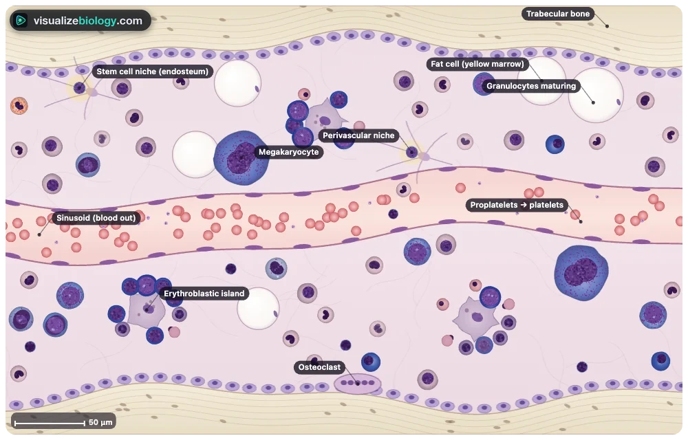
Simulate opens inside a healthy adult's red bone marrow and the clock runs (¼ day a second). The bench has four views: Body (red marrow in the skeleton by age), Marrow (a living section of marrow), Lineage (the hematopoiesis chart) and Smear (a stained bone-marrow aspirate). On the right, Cell shows the chosen cell with its parts labelled, Journey plays one cell's development, Graph plots the blood count, reticulocytes, growth factors and marrow over the days, and Life shows where blood is made from embryo to old age.
2 Patients, treatments and the clock
- Patient: pick a condition (blood donation, bleed, infection, chemotherapy, transplant, iron or B12 deficiency, kidney disease, sickle cell, aplastic anemia, AML, polycythemia vera), an age and an altitude.
- Treat: one-off actions (donate, transfuse, B12, chemotherapy, transplant, infection) and courses that stay on (iron, epoetin, filgrastim, romiplostim).
- The bar under the marrow plays and pauses the clock, sets its speed (¼ day, 1 day or 1 week a second), jumps a week or starts the patient again.
3 Reading the marrow
- Click any cell to name it and see how to recognise it. Click a niche, a macrophage, the sinusoid or a fat cell to read about it.
- The numbers of each kind of cell follow the model: chemotherapy empties the dividing stages first, a transplant refills the marrow from a few stem cells, leukemic blasts crowd out the rest, and fat cells fill an emptying marrow.
- Signals shows the growth factors arriving from the blood: EPO (gold) to the erythroblastic islands, TPO (green) to the megakaryocytes and G-CSF (blue) to the granulocytes.
- Ctrl or ⌘ + scroll zooms the marrow, the chart and the smear; drag to move around.
4 Experiment mode and the lab report
Six practicals: name the cells of a marrow smear, count the myeloid:erythroid ratio, follow the recovery after a blood donation, compare chemotherapy with and without G-CSF, time a transplant's engraftment and diagnose three mystery patients. In a practical the live blood count is hidden: take a blood test instead. Each ticks off its method as you work and ends with a conclusion question; "Lab report (PDF)" turns your notebook into a report.
5 Units and keyboard
- SI | US shows hemoglobin in g/L or g/dL and counts in ×10⁹/L or per µL; altitude in metres or feet.
- 1 Body, 2 Marrow, 3 Lineage, 4 Smear, Space play or pause, L labels, F next smear field, J journey, G graph, B blood test (Experiment), Esc clears the selection.
Hematopoiesis: how the bone marrow makes blood
Every second your bone marrow makes about 2.4 million red blood cells, over a million platelets and a million neutrophils, and it never stops. This free hematopoiesis simulator takes you inside that factory: a living section of red marrow drawn to scale, where every cell matures through its real stages and the number of each kind follows a model of the marrow. Follow one cell from the stem cell to the blood, read the full hematopoiesis chart, then give the patient a bleed, an infection, chemotherapy or a transplant and watch the marrow answer.
- Views
- Red marrow in the skeleton, a living marrow section, the hematopoiesis chart and a marrow smear
- Patients
- Blood donation, bleeding, altitude, infection, chemotherapy, transplant, iron and B12 deficiency, kidney disease, sickle cell, aplastic anemia, AML, polycythemia vera
- Standards
- NGSS HS-LS1-3 and HS-LS1-4; AP Biology; AQA GCSE 4.1.2.3 stem cells; A-level; IB B2.3
- Modes
- Simulate, Experiment (6 practicals with a PDF report) and Quiz
What is hematopoiesis?
Hematopoiesis (British haematopoiesis, also hemopoiesis) is the making of all the blood cells. It starts from hematopoietic stem cells (HSCs): about 10,000 to 20,000 active ones in an adult, each able to renew itself and to make every kind of blood cell. Their daughters, the progenitors, divide fast and commit to one family. The myeloid family makes red blood cells, platelets, granulocytes and monocytes; the lymphoid family makes B cells, T cells and natural killer cells. Together they turn out about 500 billion cells a day.
Where are blood cells made?
Before birth, blood is made first in the yolk sac (weeks 3–8), then mainly in the liver, helped by the spleen, and from about the fifth month in the bone marrow. A newborn has red, blood-making marrow in every bone. From early childhood fat replaces it in the hands and feet first, then in the shafts of the long bones, so that by about 25 the red marrow is in the trunk.
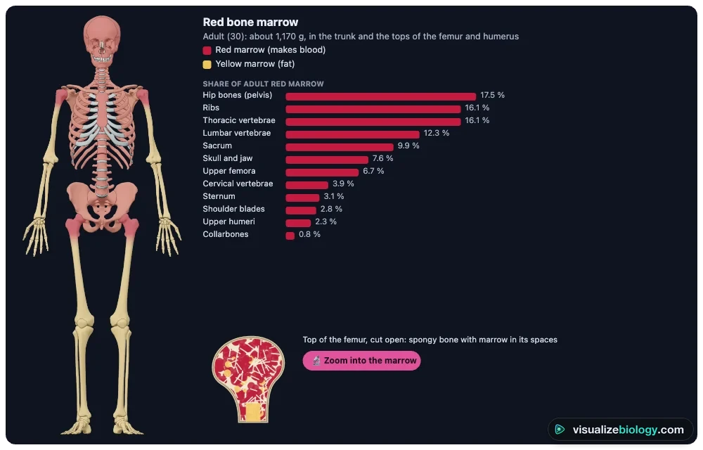
| Bone (adult) | Share of red marrow |
|---|---|
| Hip bones (pelvis) | 17.5 % |
| Ribs | 16.1 % |
| Thoracic vertebrae | 16.1 % |
| Lumbar vertebrae | 12.3 % |
| Sacrum | 9.9 % |
| Skull and jaw | 7.6 % |
| Upper femora | 6.7 % |
| Cervical vertebrae, sternum, shoulder blades, upper humeri, collarbones | 12.9 % |
The hematopoiesis chart: myeloid and lymphoid
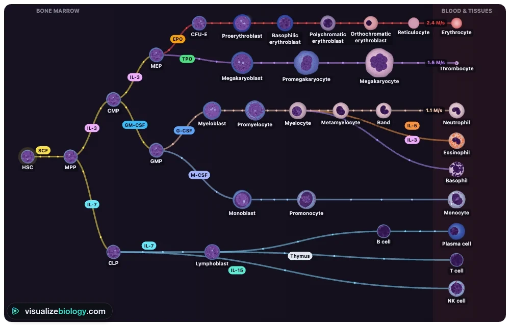
The chart below starts from one stem cell. The common myeloid progenitor splits into the megakaryocyte–erythroid progenitor (red cells and platelets) and the granulocyte–monocyte progenitor (neutrophils, eosinophils, basophils and monocytes). The common lymphoid progenitor makes B cells, which mature in the marrow, T cells, which mature in the thymus, and NK cells. Each branch is driven by a growth factor, shown on its line.
Stages of erythropoiesis
| Stage | Size | What you see | Divides? |
|---|---|---|---|
| Proerythroblast (pronormoblast) | 14–19 µm | Round nucleus, fine chromatin, nucleoli; deep blue cytoplasm | Yes |
| Basophilic erythroblast | 12–17 µm | Coarser chromatin, no nucleoli; even bluer cytoplasm | Yes |
| Polychromatic erythroblast | 10–12 µm | Checkerboard chromatin; grey-lilac cytoplasm as hemoglobin builds up | Yes, the last time |
| Orthochromatic erythroblast | 8–10 µm | Small, dense nucleus that it pushes out; pink cytoplasm | No |
| Reticulocyte | 8 µm | No nucleus; leftover RNA; 1–2 days in the marrow, 1 day in the blood | No |
| Red blood cell | 7–8 µm | Biconcave disc; lives about 120 days | No |
The whole run from proerythroblast to reticulocyte takes about 5 to 7 days. It is controlled by erythropoietin (EPO): when oxygen delivery falls (after a bleed, at altitude, in anemia) the kidneys make more EPO, more red-cell progenitors survive, and the reticulocyte count rises within a week. It is a textbook negative feedback loop.
Granulopoiesis and thrombopoiesis
Granulopoiesis runs myeloblast → promyelocyte → myelocyte → metamyelocyte → band → segmented neutrophil over about 10–14 days. Only the first three divide; the rest form a storage pool about ten times the size of the neutrophils in the blood, which an infection releases at once (bands in the blood: a "left shift"). Thrombopoiesis is different: a megakaryoblast copies its DNA without dividing, growing into a megakaryocyte of 40–100 µm that sits against a sinusoid and pushes out proplatelets, which the blood flow cuts into 1,000–3,000 platelets.
Growth factors in hematopoiesis
| Factor | Made by | Drives |
|---|---|---|
| Erythropoietin (EPO) | Kidneys, when oxygen is low | Red cells |
| Thrombopoietin (TPO) | Liver; removed by platelets | Megakaryocytes and platelets |
| G-CSF | Macrophages, endothelium; more in infection | Neutrophils |
| GM-CSF, M-CSF, IL-3 | T cells, macrophages, stroma | Granulocytes and monocytes; early myeloid cells |
| IL-5, IL-7 | T cells; marrow stroma and thymus | Eosinophils; lymphocytes |
| Stem cell factor (SCF), CXCL12 | Niche cells | Keep stem cells alive and in the marrow |
When blood making goes wrong
| Condition | What happens in the marrow | Blood count |
|---|---|---|
| Iron deficiency | Not enough iron for hemoglobin | Small, pale red cells; low reticulocytes |
| B12 or folate deficiency | DNA made too slowly: big precursors, most die | Big red cells, hypersegmented neutrophils |
| Kidney disease | Too little EPO | Normocytic anemia |
| Aplastic anemia | Stem cells lost; empty, fatty marrow | Every count low |
| Acute myeloid leukemia | 20 % or more blasts that cannot mature | Anemia, low platelets, blasts in the blood |
| Polycythemia vera | JAK2 clone grows without EPO | Too many red cells, low EPO |
Chemotherapy kills dividing cells, so neutrophils fall to a low point about 10–14 days later; a stem cell transplant replaces the whole marrow, and the donor cells' neutrophils usually appear by days 12–20. Everything here is an educational model, not medical advice.
Practicals and course links
| Practical | What you find out | Course links |
|---|---|---|
| Name the cells of a marrow smear | The stages of red and white cells | AP Bio, A-level, A&P |
| Count the M:E ratio | Erythroid hyperplasia in hemolysis | Hematology lab |
| Recovery after a blood donation | EPO feedback and reticulocytes | HS-LS1-3 |
| Chemotherapy and G-CSF | Neutropenia, nadir and growth factors | Pharmacology |
| Stem cell transplant | Engraftment of a new marrow | GCSE stem cells, HS-LS1-4 |
| Mystery marrows | Diagnosis from counts and smears | AP Bio, A-level |
Frequently asked questions
What is hematopoiesis?
Hematopoiesis (also spelled haematopoiesis, or called hemopoiesis) is the making of all blood cells: red blood cells, platelets and the white blood cells. In adults it happens in the red bone marrow, where a small pool of hematopoietic stem cells renews itself and makes about 500 billion new blood cells every day.
Where are blood cells made in the body?
In the red bone marrow. In an adult it is found in the skull, vertebrae, ribs, sternum, pelvis and the upper ends of the femur and humerus; the rest of the marrow is yellow (fat). A newborn makes blood in almost every bone. Before birth, blood is made first in the yolk sac and then mainly in the liver and spleen. T cells finish maturing in the thymus.
What are the stages of erythropoiesis?
Hematopoietic stem cell, common myeloid progenitor, megakaryocyte–erythroid progenitor, BFU-E and CFU-E, then the cells you can see on a smear: proerythroblast, basophilic erythroblast, polychromatic erythroblast, orthochromatic erythroblast (which pushes out its nucleus), reticulocyte and red blood cell. From proerythroblast to reticulocyte takes about 5 to 7 days and is driven by erythropoietin from the kidneys.
What is the difference between the myeloid and lymphoid lineages?
Both come from the hematopoietic stem cell. The common myeloid progenitor makes red blood cells, platelets (from megakaryocytes), the granulocytes (neutrophils, eosinophils, basophils) and monocytes. The common lymphoid progenitor makes the lymphocytes: B cells, which mature in the bone marrow, T cells, which mature in the thymus, and natural killer cells.
Explore related simulators
See every finished blood cell in 3D, its job and a full blood count in the Interactive Blood Cell Model. Find the bones that hold red marrow in the Interactive 3D Skeleton, follow stem cells dividing in the Mitosis Simulator, and see how the kidneys sense oxygen and make erythropoietin in the Kidney Simulator.
Sources: Keohane and others, Rodak's Hematology, 6th edition, chapters 5–7; Hoffbrand and Moss, Hoffbrand's Essential Haematology, 8th edition; Dancey and others (1976), Neutrophil kinetics in man, Journal of Clinical Investigation 58: 705–715; Kaushansky (2005), The molecular mechanisms that control thrombopoiesis, Journal of Clinical Investigation 115: 3339–3347; Cristy (1981), Active bone marrow distribution as a function of age in humans, Health Physics 41: 103–108; OpenStax Anatomy and Physiology 2e, section 18.3. The skeleton is BodyParts3D (Database Center for Life Science, CC BY 4.0). An educational model: real patients vary, and nothing here is medical advice.
Updated