
Interactive 3D Circulatory System: Every Major Artery and Vein, and How Blood Flows
Over 120 labeled arteries and veins inside a glass body, around a beating heart. Find any vessel, watch the pulse race down the arteries, then ride one red cell from the heart to the brain, a foot or the gut and back through the lungs.
Where the blood goes
Lab notebook
Every journey, activity, condition and measurement is written here. Export a CSV, or turn the session into a PDF lab report.
| # | What | Settings | Result |
|---|
Concept library
Click "New problem" to begin.
Click "Start quiz" for 5 questions on the arteries and veins of the body, the vessels, the double circulation, pulse points and blood flow.
1 Overview
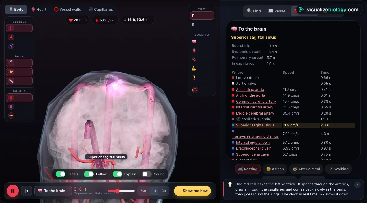
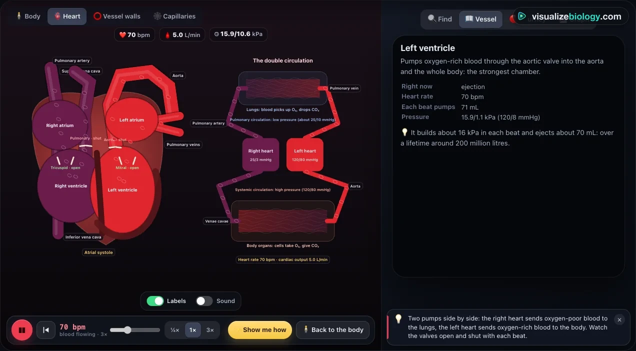
Four zoom levels sit at the top left of the view. Body is the circulatory system inside a glass body on the real skeleton, in 3D (desktop and tablet) or as front and back plates in 2D (phones). Heart shows the heart as two pumps side by side with the pulmonary and systemic circuits. Vessel walls compares an artery, a vein and a capillary in section, with a vein valve and the calf muscle pump. Capillaries follows red cells through a capillary bed giving up oxygen and making tissue fluid. The panel on the right has Find, Vessel, Journey and Graph.
2 Finding vessels
Click any vessel on the body, or type in Find: a name (femoral artery), an everyday name (wrist pulse, neck pulse, blood test vein), a region (forearm) or a job (drains the kidney). The vessel turns gold, the camera flies to it and the Vessel card shows where it comes from and goes to, what it supplies or drains, its size, its blood flow and speed right now, and its pulse point if it has one. ‹ and › walk through the vessels of the same region.
3 Journeys
Press Journeys in the bar at the bottom of the view and pick a destination: the brain, the hand, a kidney, the gut and liver, the foot, the heart muscle, the face, or through the lungs. One red cell leaves the left ventricle, runs through every named vessel on the way at its real speed, crosses the capillaries, returns to the right atrium and goes round the lungs back to the left ventricle. The clock shows the real time, and the Journey panel lists each vessel with how long the cell spends in it.
4 Activities and conditions
The Activity panel changes what the body is doing: resting, asleep, after a meal, walking, running hard, a hot day, a cold day or fight or flight. Heart rate, stroke volume, cardiac output, blood pressure and the share of blood each organ gets all change, and the organs glow with their flow. The Condition panel adds a narrowed coronary artery, carotid stenosis, high blood pressure, heart failure, blood loss, deep vein thrombosis, varicose veins or an aortic aneurysm. This is an educational model, not medical advice.
5 Experiment mode and the lab report
Six labs: name the vessels, take your pulse at rest and after exercise, radius and flow (Poiseuille), pressure and speed along the circulation, where the blood goes in exercise, and trace the double circulation. Each shows its aim, what you need and a safety note; the method ticks off as you work, then you answer the conclusion question and create a PDF lab report.
6 Quiz, units and keyboard
The practice quiz covers naming vessels, what they supply, the three kinds of vessel, the path of blood, pulse points, pressure and flow, and calculations, each with a worked solution. SI | US switches pressure between kPa and mmHg and lengths between cm and inches. Keys: 1–4 zoom levels, F find, A vessel, J journey, G graph, L labels, O rotate, Space play or pause, N back to the start, [ and ] walk the region.
The circulatory system: an interactive 3D model of the arteries, veins and heart
Your heart pumps about 5 litres of blood a minute at rest, so every drop of your blood goes round your body roughly once a minute. This free interactive 3D circulatory system shows the road it travels: over 120 named arteries and veins from the BodyParts3D anatomy database, set on a real human skeleton inside a glass body, around a heart that beats. Click any vessel to see where it runs and what it supplies, watch the pulse wave race down the arteries, and ride a single red cell from the left ventricle to the brain, a foot or the gut and back through the lungs. Change what the body is doing (a meal, a run, a hot day) and see where the blood goes.
- Vessels
- Over 120 named arteries and veins in 3D (BodyParts3D), searchable by name, region and everyday name
- Journeys
- Brain, hand, kidney, gut and liver, foot, heart muscle, face, and the lungs
- Close-ups
- The heart as a double pump; artery, vein and capillary walls; a capillary bed
- Modes
- Simulate, Experiment (6 practicals with a PDF report) and Quiz
- Price
- Free, no account or download; works on Chromebooks, tablets and phones
The major arteries of the body, labeled
Every artery in the body branches from the aorta, which leaves the left ventricle with a diameter of about 2.5–3 cm. These are the arteries students are asked to name, with what each one feeds:
| Artery | Where | Supplies |
|---|---|---|
| Aorta (ascending, arch, descending, abdominal) | From the left ventricle down in front of the spine | The whole body (all other systemic arteries) |
| Coronary arteries | From the aorta just above the aortic valve | The heart muscle |
| Common, internal and external carotid | Neck | Brain (internal), face and scalp (external) |
| Vertebral and basilar | Through the neck vertebrae into the skull | Brainstem, cerebellum, back of the brain |
| Subclavian, axillary, brachial | Under the collarbone, armpit, upper arm | Shoulder and arm |
| Radial and ulnar | Forearm | Forearm and hand |
| Celiac trunk, superior and inferior mesenteric | Front of the abdominal aorta | Stomach, liver, spleen, intestines |
| Renal arteries | Sides of the abdominal aorta | Kidneys (about 20 % of the cardiac output) |
| Common, internal and external iliac | Pelvis | Pelvic organs and the legs |
| Femoral, popliteal, tibial, dorsalis pedis | Thigh, knee, leg, foot | The leg and foot |
| Pulmonary arteries | From the right ventricle | The lungs, with oxygen-poor blood |
The major veins of the body
Veins return blood to the right atrium through two great veins. The superior vena cava brings blood from the head (through the internal jugular veins), neck and arms (subclavian, axillary, cephalic and basilic veins). The inferior vena cava brings it from the legs (femoral, popliteal and the long great saphenous vein under the skin), the pelvis (iliac veins), the kidneys (renal veins) and the liver (hepatic veins). The heart's own veins drain through the coronary sinus, and the four pulmonary veins carry oxygen-rich blood from the lungs to the left atrium. The median cubital vein in front of the elbow is the one used for blood tests.
Arteries, veins and capillaries compared
| Artery | Capillary | Vein | |
|---|---|---|---|
| Carries blood | Away from the heart | Through the tissues | Back to the heart |
| Pressure | High, pulsing (about 120/80 mmHg in the aorta) | Low, steady (about 35 falling to 15 mmHg) | Very low (under 10 mmHg) |
| Wall | Thick: elastic tissue and smooth muscle | One cell thick (endothelium only) | Thin, little muscle |
| Lumen | Narrow for its size | About 8 µm: red cells pass in single file | Wide |
| Valves | None (except the aortic and pulmonary valves) | None | Yes, in the limbs |
| Share of blood volume | About 13 % | About 5 % | About 64 % |
Double circulation: the pulmonary and systemic circuits
Mammals and birds have a double circulation: blood passes through the heart twice on each round trip. The right ventricle pumps oxygen-poor blood to the lungs at a low pressure (about 25/10 mmHg) in the pulmonary circulation; it returns oxygen-rich to the left atrium. The left ventricle then pumps it to every other organ at a high pressure (about 120/80 mmHg) in the systemic circulation. Keeping the two apart lets the body get blood fast and under pressure while the delicate lung capillaries are protected. Fish have a single circulation: the heart pumps blood through the gills and on to the body without returning, so the pressure to the body is low.
The path of blood through the body
Venae cavae → right atrium → tricuspid valve → right ventricle → pulmonary valve → pulmonary arteries → lung capillaries (oxygen in, carbon dioxide out) → pulmonary veins → left atrium → mitral (bicuspid) valve → left ventricle → aortic valve → aorta → arteries → arterioles → capillaries (oxygen out to the cells) → venules → veins → venae cavae. In the simulator a cell's trip to the brain and back takes about 20 seconds at rest, while a trip to the foot takes about a minute, mostly spent creeping back up the leg veins.
The hepatic portal system
Blood from the stomach, intestines, spleen and pancreas does not go straight back to the heart. It collects in the superior mesenteric and splenic veins, which join behind the pancreas as the hepatic portal vein, and passes through a second set of capillaries in the liver before the hepatic veins return it to the inferior vena cava. The liver gets the first look at everything absorbed from a meal: it stores glucose as glycogen, makes proteins and removes toxins and drugs. About three quarters of the liver's blood arrives this way, the rest through the hepatic artery.
Blood pressure and speed along the vessels
Pressure falls all the way round the circulation, most steeply across the arterioles, whose rings of smooth muscle set the resistance. Speed works differently: the same 5 litres a minute pass through every level, so blood moves fastest where the total cross-section is smallest. The aorta (about 2.5 cm²) carries blood at about 33 cm/s on average, but the 10 billion capillaries together have about 2500 cm², so blood crawls through them at about 0.3 mm/s, giving each red cell a second or so to exchange oxygen. It speeds up again in the veins. Blood flow through a vessel follows Poiseuille's law: flow depends on the radius to the fourth power, so halving an artery's radius cuts its flow to a sixteenth.
Pulse points
Each heartbeat sends a pressure wave down the arteries at about 5 metres per second in the aorta and 8–9 m/s in the limbs, much faster than the blood itself. Where an artery runs over a bone you can feel it: the radial pulse at the wrist, the carotid in the neck, the brachial inside the elbow, the temporal in front of the ear, the facial on the jaw, the femoral in the groin, the popliteal behind the knee, the posterior tibial behind the inner ankle and the dorsalis pedis on the top of the foot. The wave reaches the neck about 0.05 s after the heart contracts and the foot about 0.2 s after.
Blood flow during exercise
When you run, the heart rate can rise to 170 beats a minute and the stroke volume to about 120 mL, so the cardiac output climbs from 5 to around 20 L/min. Arterioles in the working muscles widen (vasodilation) while those to the gut and kidneys narrow (vasoconstriction), so the muscles' share rises from about 15 % to about 80 %. The brain keeps its 750 mL/min whatever you do. On a hot day the skin's arterioles open to lose heat; in the cold they close and fingers turn pale.
Experiments and course links
| Lab | What you find out | Course links |
|---|---|---|
| Name the vessels | Locate the major arteries and veins of the body | IGCSE 9; GCSE 4.2.2; A&P |
| Take your pulse | Heart rate at rest and after exercise; pulse points | GCSE; GCSE PE; NGSS MS-LS1-3 |
| Radius and flow | Poiseuille's law: why narrowed arteries matter | A-level; A&P |
| Pressure and speed | Pressure, total cross-section and velocity along the circulation | A-level 3.3.4.1; IB B3.2 |
| Where does the blood go? | Redistribution of cardiac output in exercise | GCSE PE; A-level PE; A&P |
| Double circulation | The order of chambers, valves and vessels | GCSE 4.2.2; IGCSE 9; NCERT 11 |
Who uses this simulator?
Middle and high school students meeting the circulatory system (NGSS, IGCSE, NCERT), GCSE students learning the double circulation and the three kinds of blood vessel, A-level and IB students studying mass transport and tissue fluid, anatomy and physiology, nursing and health science students learning the named vessels and pulse points, and teachers who want a projector-friendly model that shows blood actually moving.
Frequently asked questions
What is the difference between arteries, veins and capillaries?
Arteries carry blood away from the heart under high pressure, so they have thick walls of elastic tissue and smooth muscle and a narrow lumen; their stretch and recoil is the pulse. Veins carry blood back to the heart under low pressure, so their walls are thin, their lumen is wide and they have valves that stop blood flowing backwards; they hold about two thirds of the blood. Capillaries join them: their wall is a single layer of cells about 1 micrometre thick, so oxygen, glucose and carbon dioxide can diffuse between the blood and the tissues.
What is double circulation?
In mammals blood passes through the heart twice on each complete trip round the body. The right side of the heart pumps oxygen-poor blood through the pulmonary arteries to the lungs and back to the left atrium in the pulmonary veins: the pulmonary circulation. The left side pumps the oxygen-rich blood through the aorta to every other organ and it returns in the venae cavae to the right atrium: the systemic circulation. Because blood returns to the heart after the lungs, it can be pumped to the body at a high pressure while the lungs receive it at a low one.
What is the path of blood through the body?
Starting in the body: venae cavae, right atrium, tricuspid valve, right ventricle, pulmonary valve, pulmonary trunk and arteries, capillaries of the lungs (where the blood picks up oxygen), pulmonary veins, left atrium, mitral (bicuspid) valve, left ventricle, aortic valve, aorta, arteries, arterioles, capillaries of an organ (where it gives up oxygen), venules, veins and back to the venae cavae. A round trip takes about a minute at rest.
Where can you feel your pulse?
Wherever an artery runs close to the skin over a bone or firm tissue: the radial artery at the thumb side of the wrist, the carotid artery in the neck beside the windpipe, the brachial artery inside the elbow, the superficial temporal artery in front of the ear, the facial artery on the lower edge of the jaw, the femoral artery in the groin, the popliteal artery behind the knee, the posterior tibial artery behind the inner ankle and the dorsalis pedis artery on the top of the foot.
Is this 3D circulatory system free, and does it need a login?
Yes, it is free. There is no account, sign-up, download or plug-in: it runs in any modern browser on a Chromebook, laptop, tablet or phone, and nothing a student does is sent anywhere. Teachers can share a link or a classroom QR code from the share bar. It is an educational model, not medical advice.
Explore related simulators
The pump itself, with the cardiac cycle, the Wiggers diagram and the ECG, is in the Heart Simulator: Blood Flow & Cardiac Cycle. Measure the pressure these arteries carry with the Manual Blood Pressure Apparatus Simulator, and meet the cells the blood carries in the Interactive Blood Cell Model. The muscles that pump the leg veins are in the Interactive 3D Muscular System, and the bones the vessels run beside in the Interactive 3D Skeleton.
Sources: OpenStax Anatomy and Physiology 2e, chapters 19–20; OpenStax Biology 2e, chapter 40; Hall & Hall, Guyton and Hall Textbook of Medical Physiology, 14th edition, chapters 14–21; Burton, Physiology and Biophysics of the Circulation (1965); Gray's Anatomy, 42nd edition; The Reference Values for Arterial Stiffness' Collaboration, Eur. Heart J. 31 (2010). 3D vessels, heart, organs, skin and bones: BodyParts3D, © The Database Center for Life Science, licensed under CC BY 4.0 (simplified, with centre lines added; the external carotid, facial and temporal arteries, external jugular vein, dural venous sinuses and fibular artery are drawn along landmarks). Educational model, not medical advice.
Updated