
Liver Function Simulator: See What the Liver Does, in 3D
Turn a real human liver, open its segments and vessels, then zoom into a living lobule and one hepatocyte. Feed, fast, drink or overdose the patient and watch glycogen, bile, bilirubin, alcohol, drugs and the blood tests change.
The liver now
Lab notebook
Every patient you choose, every meal, drink or treatment and every test is written here. Export a CSV, or turn the session into a PDF lab report.
| # | Patient | What | Result | Value |
|---|
Concept library
Click "New problem" to begin.
Click "Start quiz" for 5 questions on the liver's anatomy, lobule, functions, jaundice, alcohol and drugs, and its blood tests.
1 Overview
Simulate opens on a healthy adult's liver with the clock running (10 minutes a second, meals at 8:00, 13:00 and 19:00). The bench has three views: Liver (the real organ in 2D or 3D), Lobule (a living lobule under the microscope) and Cell (one hepatocyte). On the right, At work shows the liver's seven functions live, Graph plots the blood tests over time and Patient shows the eyes, urine and stool. "The liver now" lists the liver function tests and whatever you clicked.
2 The liver view: looks, cameras and 3D
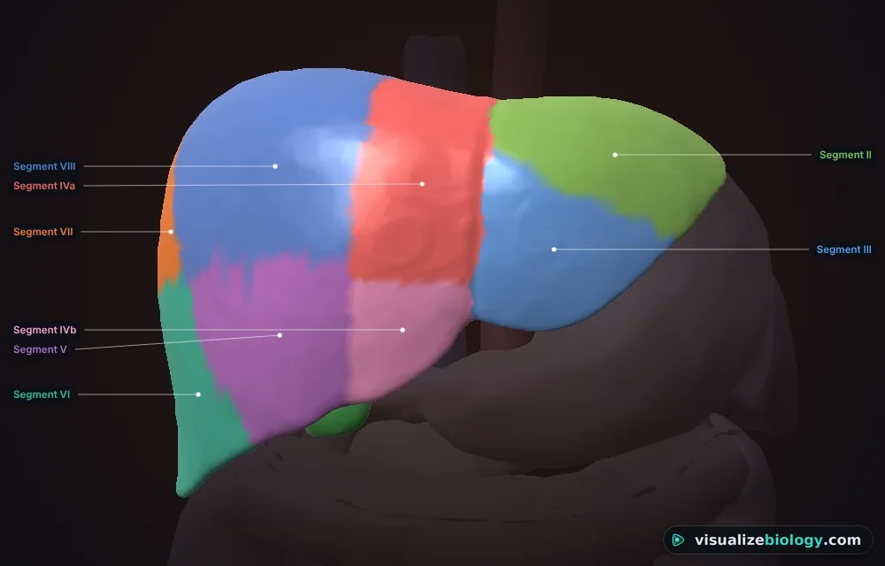
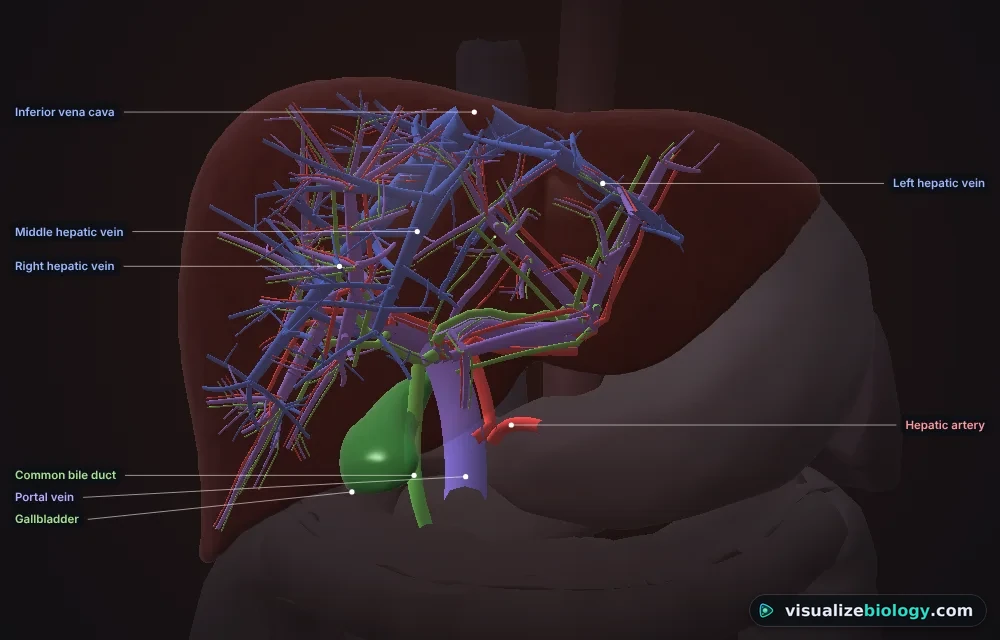
- Look: Surface, Segments (Couinaud I–VIII, with IVb), Lobes (right, left, caudate, quadrate) or Vessels (the liver turns to glass over the portal vein, hepatic artery, bile ducts and hepatic veins, with blood and bile flowing).
- Camera (2D): from the front, from below (the visceral surface) or from the right side. In 3D drag to turn, scroll with Ctrl/⌘ or pinch to zoom, and press Explode to pull the segments apart.
- Click any segment, lobe, vessel or the gallbladder to read about it. The surface changes with the patient: pale and yellow in fatty liver, nodular in cirrhosis, mottled after an overdose.
3 The lobule and the hepatocyte
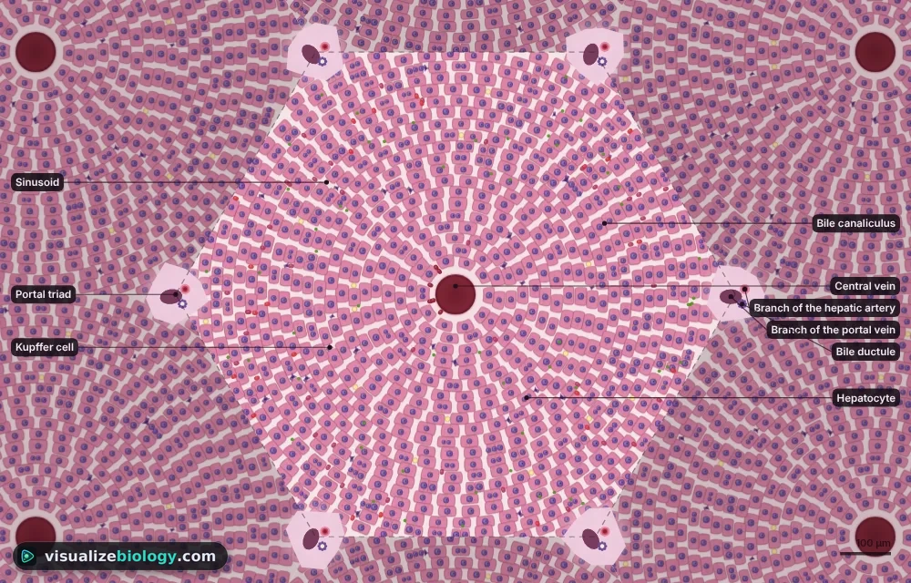
- Lobule: blood runs from the portal triads at the corners to the central vein; bile runs the other way. Zones colours zones 1–3; PAS stains glycogen magenta (fed vs fasting). Ctrl/⌘ + scroll zooms, drag pans. Click a hepatocyte to open it.
- Cell: pick a function (glucose, ammonia, bilirubin, bile, alcohol, drugs, proteins). Molecules travel at the model's rate and the numbered steps light up as they happen.
4 Patients, actions and the clock
- Patient: healthy, fatty liver, alcohol-related liver disease, acute hepatitis, cirrhosis, a gallstone in the bile duct, hemolytic anemia, Gilbert syndrome, newborn jaundice or an acetaminophen overdose; male or female body.
- Do: four kinds of meal, a standard drink (14 g of alcohol), acetaminophen 1 g or an overdose, N-acetylcysteine, vitamin K, ERCP (removes the stone), and the courses fasting, lactulose and phototherapy.
- The bar under the liver plays and pauses the clock, sets its speed (1 minute to 1 day a second), jumps 6 hours or starts the patient again.
5 Experiment mode and the lab report
Five practicals: label the liver and lobule, catalase in liver at different temperatures, the liver function test detective (four mystery patients), alcohol elimination (Widmark) and treating an acetaminophen overdose. In a practical the live blood tests are hidden: take a test from the Lab panel. Each ticks off its method as you work and ends with a conclusion question; "Lab report (PDF)" turns your notebook into a report.
6 Units and keyboard
- SI | US: bilirubin µmol/L or mg/dL, glucose mmol/L or mg/dL, urea mmol/L or BUN mg/dL, ammonia µmol/L or µg/dL, albumin g/L or g/dL, blood alcohol g/L or %, body mass kg or lb, temperature °C or °F.
- 1 Liver, 2 Lobule, 3 Cell, Space play or pause, L labels, G graph, W at work, P patient, Esc clears the selection.
The liver: what it does and how it works
The liver is the body's biggest internal organ (about 1.5 kg) and its chemical factory: it stores glucose, makes bile, clears bilirubin, turns toxic ammonia into urea, breaks down alcohol and medicines, and makes most of the proteins in your blood. This free liver function simulator lets you see all of it: a real human liver in 3D, a living lobule under the microscope and a single hepatocyte at work, all driven by one model of the liver that responds to meals, fasting, alcohol, drugs and disease.
- Views
- The real liver in 2D and 3D (surface, segments, lobes, vessels), a living lobule and one hepatocyte
- Patients
- Fatty liver, alcohol-related liver disease, hepatitis, cirrhosis, gallstones, hemolysis, Gilbert syndrome, newborn jaundice, acetaminophen overdose
- Standards
- NGSS HS-LS1-2 and HS-LS1-3; AP Biology; AQA GCSE 4.2.2 and 4.5.3; A-level deamination and the urea cycle; A&P
- Modes
- Simulate, Experiment (5 practicals with a PDF report) and Quiz
The functions of the liver
| Function | What the liver does | In numbers |
|---|---|---|
| Glucose store | Stores glucose as glycogen after a meal (insulin); releases it, then makes new glucose (gluconeogenesis), when fasting (glucagon) | About 100 g of glycogen, enough for a day |
| Ammonia → urea | Deaminates amino acids and turns the toxic ammonia into urea in the urea cycle | About 30 g of urea a day |
| Bile | Makes bile salts from cholesterol; bile emulsifies fats in the small intestine | About 600 mL a day |
| Bilirubin | Conjugates bilirubin from old red cells and excretes it in bile | About 300 mg a day |
| Detoxification | Breaks down alcohol, drugs and hormones (CYP450 enzymes, glucuronidation, glutathione) | About one drink an hour |
| Plasma proteins | Makes albumin and clotting factors (with vitamin K) | About 12 g of albumin a day |
| Storage and defense | Stores vitamins A, D and B12, iron and copper; Kupffer cells remove bacteria and old red cells | Vitamin A for months |
Liver anatomy: lobes, segments and blood supply
From the front the falciform ligament divides a big right lobe from a smaller left lobe; from below you also see the caudate and quadrate lobes, the gallbladder and the porta hepatis, where vessels enter and the bile ducts leave. Surgeons use the eight Couinaud segments instead: each has its own branch of the portal vein, hepatic artery and bile duct, and the hepatic veins run between them, so one segment can be removed without harming the rest. The liver has a dual blood supply: about 75 % of its blood comes from the hepatic portal vein, full of nutrients from the gut, and 25 % from the hepatic artery, rich in oxygen. All of it leaves by the hepatic veins into the inferior vena cava.
The liver lobule and its zones
Under the microscope the liver is made of hexagonal lobules about 1 mm across. At each corner is a portal triad (a branch of the portal vein, a branch of the hepatic artery and a bile ductule); in the middle is the central vein. Plates of hepatocytes one cell thick radiate between them, with leaky capillaries called sinusoids in between, lined by Kupffer cells. Blood flows inward from the triads to the central vein, while bile flows outward in tiny canaliculi between the hepatocytes. Cells near the triads (zone 1) get the most oxygen and do most gluconeogenesis and urea making; cells near the central vein (zone 3) do most drug metabolism, which is why acetaminophen damages them first.
Bilirubin and the three types of jaundice
Jaundice is yellow skin and eyes from too much bilirubin (over about 50 µmol/L, 3 mg/dL). Where the problem lies decides which bilirubin rises:
| Type | Cause | Blood | Urine | Stool |
|---|---|---|---|---|
| Pre-hepatic | Too many red cells broken down (hemolysis) | Unconjugated bilirubin high | No bilirubin, urobilinogen high | Dark |
| Hepatic | Liver cells damaged (hepatitis, cirrhosis); Gilbert syndrome; newborns | Both kinds high, ALT high | Bilirubin present | Normal or pale |
| Post-hepatic | Bile duct blocked (gallstone, tumor) | Conjugated bilirubin high, ALP and GGT high | Dark, bilirubin +++, no urobilinogen | Pale (clay-colored) |
Liver function tests
| Test | Normal | High or low means |
|---|---|---|
| ALT, AST | Under about 40 U/L | Liver cells dying; AST over twice ALT suggests alcohol |
| ALP, GGT | ALP 44–147, GGT under 61 U/L | Bile not flowing (cholestasis); GGT also rises with alcohol |
| Bilirubin | 3–21 µmol/L (0.2–1.2 mg/dL) | Jaundice (see above) |
| Albumin | 35–50 g/L (3.5–5.0 g/dL) | Low when the liver cannot make proteins (cirrhosis) |
| INR | About 1.0 | High when clotting factors are not made (liver failure) or vitamin K is missing |
How the liver breaks down alcohol and acetaminophen
Alcohol is oxidised by alcohol dehydrogenase to acetaldehyde, then by ALDH2 to acetate. The enzyme is saturated even at low levels, so the liver clears a fixed amount, about 0.15 g/L of blood alcohol an hour (roughly one drink an hour), whatever you drink; coffee and cold showers do not speed it up. The extra NADH blocks gluconeogenesis and builds fat, which is why heavy drinking causes fatty liver. Acetaminophen (paracetamol) is mostly glucuronidated and sulfated, but a few per cent become NAPQI, a toxic molecule that glutathione neutralises. In an overdose those pathways saturate, glutathione runs out and NAPQI kills zone-3 hepatocytes over the next one to three days. N-acetylcysteine refills glutathione and works best within 8 hours. Everything here is an educational model, not medical advice: anyone who has taken too much acetaminophen needs emergency care at once.
Practicals and course links
| Practical | What you find out | Course links |
|---|---|---|
| Label the liver and its lobule | Lobes, vessels, triads, central vein | GCSE, A&P |
| Catalase in liver | Enzyme activity and temperature | GCSE and A-level enzymes |
| Liver function test detective | The three jaundices and cirrhosis | A-level, AP Bio, A&P |
| Alcohol elimination | Widmark's formula and zero-order kinetics | A-level, AP Bio |
| Treating an acetaminophen overdose | NAPQI, glutathione and the antidote | A-level, pharmacology |
Frequently asked questions
What are the main functions of the liver?
The liver stores glucose as glycogen and releases it between meals, turns ammonia into urea, makes about 600 mL of bile a day, conjugates and excretes bilirubin, breaks down alcohol, drugs and hormones, makes albumin and clotting factors, and stores vitamins A, D and B12, iron and copper. Its Kupffer cells also remove bacteria and old red blood cells.
What are the three types of jaundice?
Pre-hepatic jaundice comes from too much red-cell breakdown (hemolysis): unconjugated bilirubin rises and the urine has no bilirubin. Hepatic jaundice comes from damaged liver cells (hepatitis, cirrhosis): both kinds of bilirubin rise and liver enzymes are high. Post-hepatic (obstructive) jaundice comes from a blocked bile duct, often a gallstone: conjugated bilirubin, ALP and GGT rise, the urine is dark and the stools are pale.
What is a liver lobule?
A liver lobule is the liver's small hexagonal unit, about 1 mm across. Portal triads (a branch of the portal vein, a branch of the hepatic artery and a bile ductule) sit at its corners and a central vein at its middle. Plates of hepatocytes radiate between them with sinusoids in between: blood flows inward to the central vein and bile flows outward to the bile ductules.
How fast does the liver remove alcohol?
About 0.15 g/L of blood alcohol an hour (0.015 % BAC an hour), roughly one standard drink an hour. Alcohol dehydrogenase is saturated even at low levels, so the rate stays about the same however much is drunk, and nothing such as coffee or a cold shower speeds it up.
Explore related simulators
Follow a meal from mouth to colon, with bile emulsifying fat, in the Human Digestive System Simulator. See how the liver, pancreas and muscles keep blood sugar steady in the Blood Glucose Regulation Simulator, how the kidneys excrete the urea the liver makes in the Kidney Simulator, and the red blood cells whose hemoglobin becomes bilirubin in the Interactive Blood Cell Model. Measure catalase and other enzymes in the Enzyme Kinetics Virtual Lab.
Sources: Hall and Hall, Guyton and Hall Textbook of Medical Physiology, 14th edition, chapter 71; Boron and Boulpaep, Medical Physiology, 3rd edition, chapter 46; Couinaud (1957), Le foie: études anatomiques et chirurgicales; Rumack and Matthew (1975), Acetaminophen poisoning and toxicity, Pediatrics 55: 871–876; Widmark (1932); OpenStax Anatomy and Physiology 2e, section 23.6. The 3D liver, gallbladder, pancreas and surrounding organs are BodyParts3D 4.0 (© DBCLS, CC BY 4.0; Mitsuhashi and others 2009); the vessel trees inside the liver are drawn by the simulator from its segments.
Updated